
Anatomy of Sports Injuries: How to Identify and Prevent Common Damage
Aug 31, 2025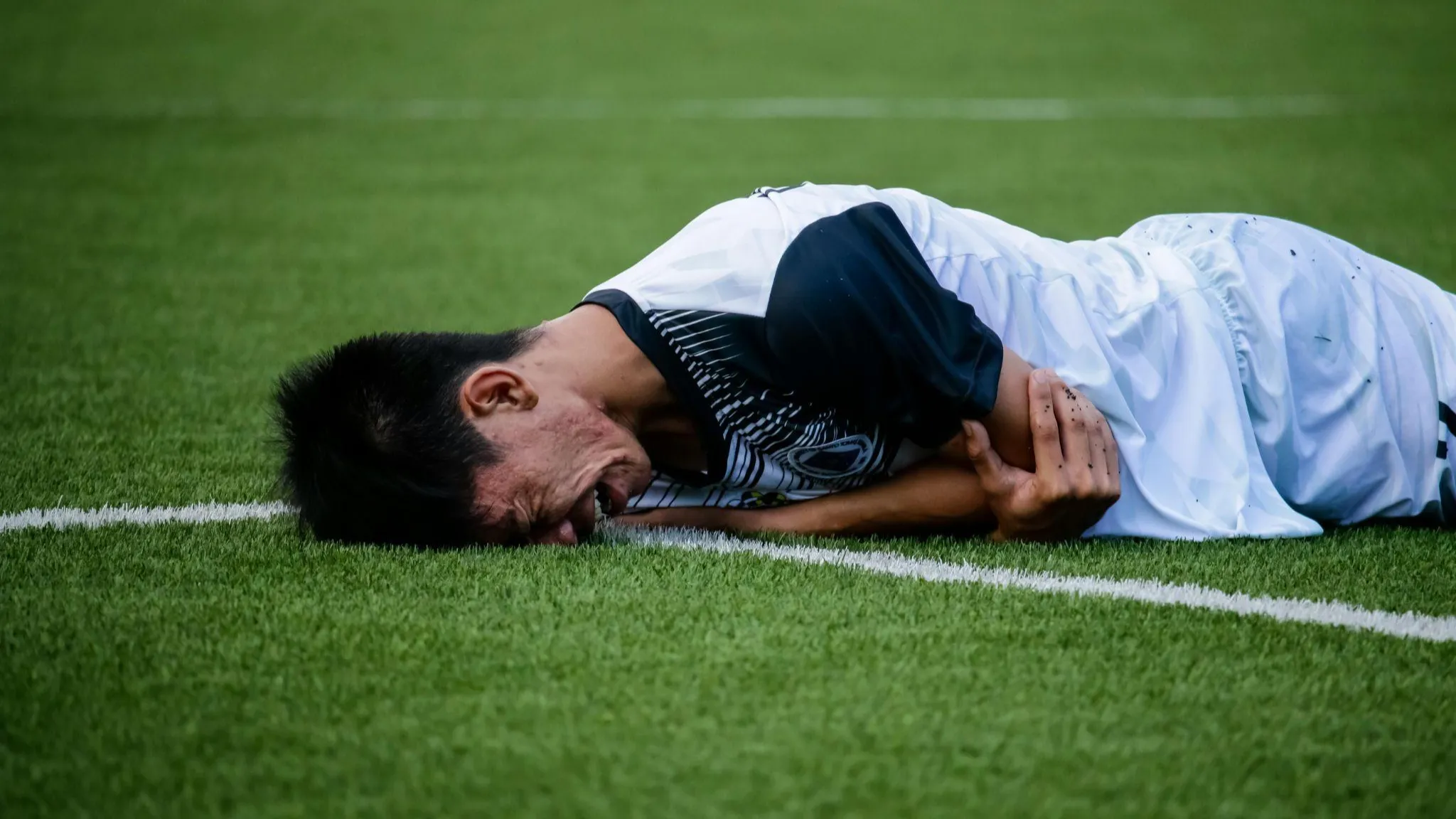
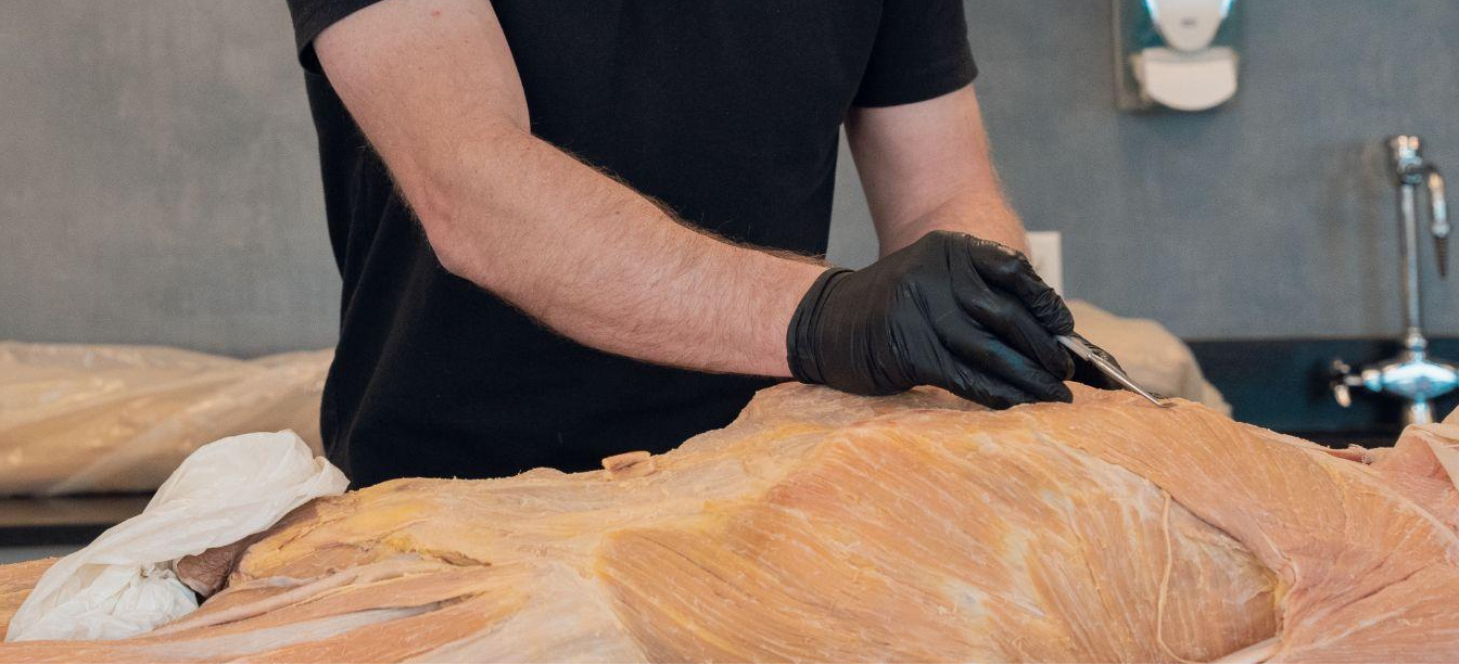
Sports injuries are more than just setbacks; they’re windows into the intricate design of the human body. At the Institute of Human Anatomy, we believe that understanding the “why” behind injuries empowers athletes, trainers, and anatomy enthusiasts to train smarter and recover stronger. In this guide, we’ll dissect the anatomy of three common injuries, explore their biomechanical causes, and provide actionable prevention strategies rooted in science.
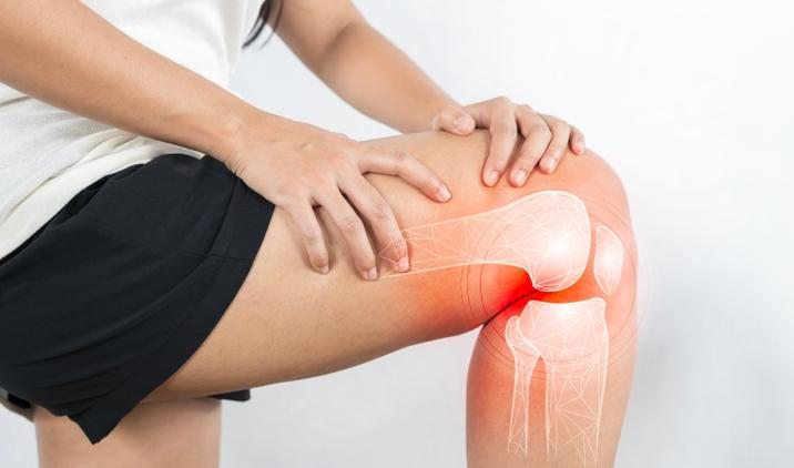
1. ACL Tears: The Knee's Fragile Guardian
Anatomy Deep Dive
The anterior cruciate ligament (ACL) is one of four major ligaments in the knee, but its role is unique. Originating from the femur’s posterior aspect and attaching to the tibia’s anterior intercondylar area, it resists forward tibial translation and rotational forces. Unlike the medial collateral ligament (MCL), which can often heal on its own, the ACL’s poor blood supply makes natural recovery nearly impossible.
Why ACL Injuries Occur
- Biomechanical Weakness: The ACL is most vulnerable when the knee is slightly bent (20–30°), a common position during cutting motions in soccer or basketball.
- Muscle Imbalances: Overdeveloped quadriceps paired with weak hamstrings increase strain on the ACL.
- Hormonal Factors: suggest estrogen fluctuations may temporarily loosen ligaments, raising women’s ACL injury risk by 2–8x compared to men.
Case Study: The “Non-Contact” Tear
About 70% of ACL tears happen without direct contact. In FIFA athletes, this often occurs during sudden stops, pivots, or awkward landings. Similarly, imagine a volleyball player landing from a jump with knees collapsing inward (valgus collapse). This misalignment places up to six times body weight on the ACL, exceeding its 500N tensile strength.
Prevention Strategies for Volleyball Athletes
Neuromuscular Training: Volleyball-specific programs teach proper landing mechanics, deceleration, and hip-knee alignment, reducing ACL injury risk.
Hamstring Strengthening: Exercises like Nordic hamstring curls build strength in muscles that protect the ACL during explosive jumps and landings.
Proprioceptive Drills: Single-leg balance and controlled landing drills improve joint stability and body awareness on the court.
Rehab Connection
Should surgery be required, long-term recovery depends on following milestone-based benchmarks rather than an arbitrary calendar date. For a complete breakdown of the exercises and tests needed to safely rebuild your knee, read our comprehensive guide: ACL Rehab Protocols: Evidence-Based Practices.
For anatomy enthusiasts, our Foundations of Human Anatomy course covers joint mechanics in detail.
2. Rotator Cuff Injuries: Decoding Shoulder Stability
Anatomy Deep Dive
The rotator cuff isn’t a single structure; it’s a quartet of muscles (supraspinatus, infraspinatus, teres minor, subscapularis) that form a “cuff” around the humeral head. Their tendons fuse into a continuous sheath, making isolated injuries rare. The supraspinatus is most vulnerable due to its position under the acromion.
Why Rotator Cuff Damage Happens
- Impingement Syndrome: Repeated overhead motions (e.g., baseball pitching) cause the supraspinatus tendon to rub against the acromion, leading to inflammation.
- Tendon Degeneration: After age 40, reduced collagen synthesis increases tear risk, even without trauma.
- Scapular Dyskinesis: Weak serratus anterior muscles let the scapula “wing,” altering shoulder mechanics.
Case Study: The Swimmer’s Shoulder
Competitive swimmers perform ~4,000 strokes per session. Over time, lax anterior ligaments and tight posterior capsules create instability, leading to partial-thickness tears.
Prevention Strategies
- Scapular Stabilization: Prone Y-T-W exercises activate the lower trapezius.
- Load Management: Limit throws or serves to 80/week for adolescent athletes.
Rehab Connection
Eccentric exercises lowering a weight slowly) stimulate tendon remodeling.
For professionals, our courses explore connective tissue biology and principles applicable to tendons.
3. Tendonitis: When Overuse Outpaces Healing
Anatomy Deep Dive
Tendons transmit muscle force to bone via organized collagen fibers. Unlike muscles, they have a 7x lower metabolic rate, slowing recovery. Chronic overuse disrupts collagen alignment, causing tendinosis (degeneration) masked as “tendonitis” (inflammation).
Common Types
- Achilles Tendonitis: Often linked to calf tightness and excessive uphill running.
- Lateral Epicondylitis (Tennis Elbow): Involves microtears in the extensor carpi radialis brevis tendon.
Why Tendonitis Persists
- Failed Healing Response:
Tendonitis often becomes chronic because of a poor healing cycle. Repeated overuse depletes tenocyte cells, the specialized cells responsible for producing collagen and maintaining healthy tendon structure. As these cells diminish, the tendon struggles to repair itself properly, leading to ongoing pain and weakness. - Neovascularization:
In chronic tendonitis, the body responds to damage by growing new, abnormal blood vessels into the injured tendon, a process called neovascularization. Along with these vessels come new nerve fibers, which increase the sensitivity of the area, causing persistent discomfort even during light activity.
Prevention and Management Strategies
- Load Monitoring:
To prevent tendon overload, use tools like the Acute:Chronic Workload Ratio (ACWR), which compares an athlete’s recent training load to their longer-term average. Research suggests keeping weekly increases in workload below 1.2 times the previous average helps reduce injury risk. - Isometric Holds:
Isometric exercises, like 45-second mid-range holds, are effective for reducing tendon pain without placing excessive strain on the tendon. For example, a wall sit is a common isometric hold for managing patellar tendonitis, helping to strengthen surrounding muscles and improve tendon load tolerance. - Footwear Matters:
In activities involving the lower limbs, footwear can significantly affect tendon strain. Shoes with a heel drop (the difference between heel and forefoot height) greater than 10mm have been shown to increase strain on the Achilles tendon by up to 18%. Selecting appropriate footwear with a moderate heel drop can reduce unnecessary stress and support tendon recovery.
Rehab Connection
Heavy slow resistance (HSR) training, 3 sets of 15 reps at 70% 1RM, rebuilds collagen density.
For a complete guide on how to safely structure these loading intervals and prevent minor microtears from turning into chronic degeneration, read our full article: How Overuse Damages Tendons and Ligaments.
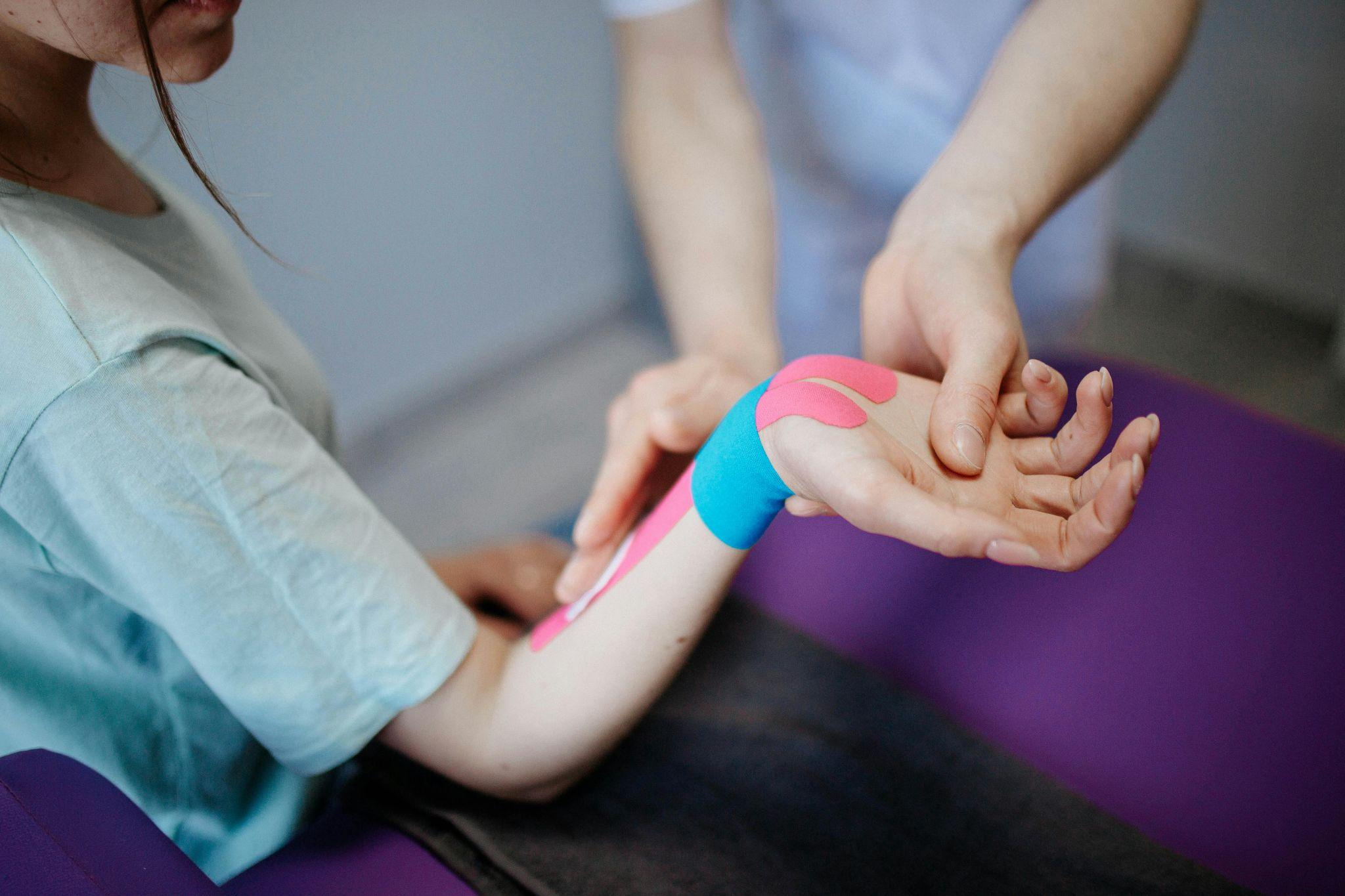
4. Injury Rehabilitation: A 4-Phase Framework
A structured recovery plan ensures tendons and joints heal progressively, reducing reinjury risk. Here's how each phase works, what to do, and why it matters:
Phase 1: Protect & Reduce Inflammation (Days 0–7)
Goal: Minimize tissue damage and control inflammation.
- POLICE Protocol: Protect the area, apply Optimal Loading (gentle, pain-free movement), Ice, Compression, and Elevation. This modern update replaces the outdated RICE protocol.
- Pulsed Ultrasound: Stimulates fibroblast activity in superficial tendons, promoting early tissue repair.
Phase 2: Restore Mobility (Weeks 2–4)
Goal: Improve joint range of motion and reduce stiffness.
- Grade III Joint Mobilizations: Manual therapy techniques where a clinician applies large-amplitude, end-range oscillations to the joint capsule. This helps break up adhesions and improve mobility after immobility.
Example: Mobilizing the knee after ACL surgery to regain flexion. - Foam Rolling: Self-myofascial release to reduce muscle adhesions and improve tendon glide. Focus areas might include quadriceps, hamstrings, and calves.
- Mobility Exercises:
- Heel slides for knee flexion
- Pendulum swings for shoulder stiffness
- Ankle alphabet to restore ankle mobility
Phase 3: Rebuild Strength (Weeks 4–12)
Goal: Restore muscle strength and tendon load tolerance.
- Isokinetic Machines: Provide resistance matched to the effort throughout the movement, making them ideal for controlled strengthening (e.g., knee extensions for ACL rehab).
- Blood Flow Restriction (BFR) Training: Uses cuffs to partially restrict blood flow during low-load exercises, allowing muscle growth and strength gains at just 20-30% of maximal loads.
- Strength Exercises:
- Leg press and squats (progressing from bodyweight to weighted)
- Hamstring curls
- Isometric holds for the affected tendon (e.g., wall sits for patellar tendonitis)
Phase 4: Return to Sport (Months 3-6)
Goal: Restore sport-specific movement patterns, balance, and confidence.
- Biodex Balance Tests: Quantify proprioceptive control by challenging stability on force plates or dynamic surfaces.
- Sport-Specific Drills: Mimic the physical demands of your sport.
Examples: - Cutting drills for soccer and football
- Plyometric jumps and landings for volleyball
- Agility ladder work for basketball
For evidence-based protocols, the National Institute of Arthritis and Musculoskeletal and Skin Diseases (NIAMS) offers free guidelines.
5. Prevention Through Biomechanical Mastery
Warm-Up Science:
A proper dynamic warm-up does more than just loosen muscles; it prepares the entire neuromuscular system for performance. Increasing muscle temperature by 2-3°C reduces viscous resistance within muscle fibers, allowing them to contract more efficiently and with less risk of strain. Additionally, warm-ups enhance joint range of motion, improve nerve conduction speed, and elevate oxygen delivery to working tissues.
Key Benefits:
- Reduces muscle stiffness and improves elasticity
- Enhances coordination and reaction times
- Prepares tendons and ligaments for load-bearing movements
Recommended Movements:
- Leg Swings: Mobilize the hip flexors and hamstrings, promoting dynamic flexibility in the lower body.
- Inchworms: Activate the posterior chain (hamstrings, glutes, lower back) and improve core stability through controlled trunk flexion and extension.
6. How the Institute of Human Anatomy Elevates Your Knowledge
Some sport injuries are random misfortunes but biomechanical puzzles rooted in anatomy. Understanding the why behind ACL tears, rotator cuff strain, or tendonitis empowers you to train smarter. Prevention lies in respecting the body’s design: optimize movement patterns, prioritize recovery, and address weaknesses before they escalate.
At the Institute of Human Anatomy, we transform complex anatomy into actionable insights. Explore our courses to decode injury mechanisms or dive into rehabilitation strategies. Remember, the best injury prevention is knowledge of your body’s limits, its strengths, and the science that bridges them. Stay curious, stay resilient.

