
Hippocampus and Aging: Memory Decline Explained
Jan 17, 2026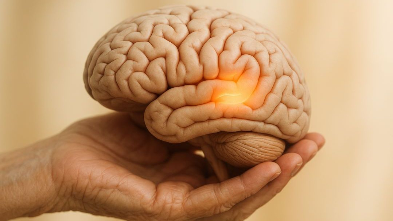
Ever forget where you parked or why you entered a room? These common memory lapses often involve the hippocampus, a brain region crucial for forming and recalling memories. As we age, the hippocampus undergoes physical and biological changes that can make learning new information or recalling recent events more challenging. Here's what you need to know:
- What the hippocampus does: It helps create episodic memories (personal experiences) and spatial maps (like remembering locations).
- Aging effects: Gradual shrinkage, reduced neurogenesis (new brain cells), and weaker neural connections lead to slower recall and occasional forgetfulness.
- Normal vs. disease: Mild lapses are typical with age, but frequent memory loss, confusion, or difficulty managing tasks could signal conditions like Alzheimer's.
- Prevention tips: Regular exercise, mental stimulation, quality sleep, and stress management can help support hippocampal health.
Understanding these changes can help distinguish normal aging from more serious memory issues while offering ways to maintain brain health as you age.
Relating the age related decline in long term memory to hippocampal structure and function
How Aging Affects the Hippocampus
As mentioned earlier, gradual changes in the brain help explain common memory lapses that come with age. The hippocampus, a key region for memory and learning, undergoes physical and cellular changes over decades. These shifts often make it harder for older adults to learn new information or quickly recall recent events. Recognizing these changes can help differentiate normal aging from more concerning conditions. The structural and biological transformations in the hippocampus set the stage for memory challenges that will be discussed further.
➡️For a foundational guide to how the entire nervous system operates, read our blog on How the Nervous System Works: A Beginner's Guide to Brain and Nerve Function
What Structural Changes Occur in the Aging Hippocampus?
Hippocampal atrophy - most pronounced in areas like the dentate gyrus and CA3 - has been a consistent finding in MRI and long-term studies. These regions show marked volume reduction and synaptic loss. Dendritic spines, which are critical for neuron-to-neuron communication, lose density and complexity with age, making it harder for the hippocampus to process and store new information.
The perforant path, the main route for information entering the hippocampus, is particularly vulnerable. Studies show that its structure degrades over time due to its long projections and high metabolic demands. This breakdown reduces both the quality and amount of information reaching the hippocampus, leading to difficulties in forming new memories.
Another key change is the decline in neurogenesis. The dentate gyrus, which generates new neurons throughout life to maintain the hippocampus's adaptability, slows down this process with age. This reduction in neurogenesis contributes to the hippocampus shrinking over time.
These structural changes also affect how the hippocampus processes information. Aging often shifts the balance between pattern separation - the ability to distinguish between similar experiences - and pattern completion, which involves recalling old memories from partial cues. As a result, encoding new memories becomes more challenging, while established memories remain relatively intact.
What Biological Mechanisms Drive These Changes?
Several interconnected biological processes contribute to the structural changes in the hippocampus, creating a cascade of stress on its cells.
One major factor is oxidative stress. Over time, cells accumulate reactive oxygen species, which damage proteins, lipids, and DNA. This damage can disrupt synaptic connections and reduce neurogenesis, speeding up cognitive decline.
Neuroinflammation adds to the problem. The hippocampus experiences an increase in inflammatory markers and chronic low-level immune activation. This persistent inflammation interferes with synaptic plasticity and reduces levels of brain-derived neurotrophic factor (BDNF), a growth factor essential for memory and learning.
Changes in synaptic plasticity also play a role. Aging brains often show reduced long-term potentiation (LTP) - a process that strengthens synaptic connections - and a greater tendency toward long-term depression (LTD), which weakens them. These shifts in synaptic plasticity have been closely linked to the memory difficulties seen in older adults.
Additionally, neurotransmitter systems that regulate hippocampal activity decline with age. For example, the locus coeruleus, which provides noradrenaline to the dentate gyrus, becomes less active, leading to imbalances in hippocampal network function.
Vascular changes further contribute to the decline. Blood vessels lose elasticity, new blood vessel formation slows, and the blood-brain barrier may weaken. These changes reduce the delivery of oxygen and nutrients that the hippocampus needs to function effectively.
Finally, epigenetic modifications play a significant role. Increased activity of histone deacetylases (HDACs) can suppress genes involved in synaptic plasticity and memory formation. Interestingly, studies in animal models suggest that reversing these epigenetic changes with HDAC inhibitors could restore some cognitive functions, offering potential therapeutic possibilities.
All these biological processes work together to gradually weaken the hippocampus. However, there’s some encouraging news: many of these changes - and their effects on memory - can be influenced by lifestyle choices, a topic that will be explored in upcoming sections.
Memory Decline and the Aging Hippocampus
As we age, changes in the hippocampus can directly impact memory, leading to challenges that range from mild lapses to more serious impairments. Knowing which memory functions are most affected and recognizing when these changes deviate from normal aging can help individuals seek timely and appropriate care.
What Memory Functions Are Most Affected by Hippocampal Aging?
The hippocampus plays a central role in memory, but not all types of memory are equally impacted by aging. Among the first to show decline is episodic memory - the ability to recall personal experiences and events. This is the type of memory that helps you remember what you had for breakfast or a conversation from last week. Because the hippocampus is critical for forming these memories, age-related changes in its structure can make this process more challenging over time.
Spatial memory, which helps us navigate and remember locations, also declines noticeably. Older adults might struggle to find their way in new environments or forget where they parked the car. Acting like an internal GPS, the hippocampus becomes less reliable as its structure degrades. Studies in both humans and animals consistently show that older adults have more difficulty creating detailed memories of places, even as their general knowledge and vocabulary remain unaffected.
Another area that becomes vulnerable is contextual or source memory - the ability to recall the circumstances of how, when, or where something was learned. For example, someone might remember a fact but forget whether they heard it on the news, read it in a book, or learned it from a friend. This type of memory heavily depends on the hippocampus to piece together the details of an experience.
Pattern separation, the ability to distinguish between similar experiences or locations, also weakens with age. Older adults are more likely to confuse where something happened or mix up similar events. Research tasks requiring participants to differentiate between nearly identical images consistently show poorer performance in older adults, linked to changes in specific hippocampal regions like the dentate gyrus and CA3.
Another hallmark of hippocampal aging is slower learning. Older adults often need extra time or more repetitions to retain new information. On memory tests, they may perform worse on delayed recall tasks, such as remembering word lists or stories, which rely heavily on the hippocampus. In daily life, this might translate to relying more on written notes, calendars, or phone reminders to keep track of new information. However, long-held memories and skills, such as riding a bike or typing, often remain intact.
The gradual decline in hippocampal function can lead to everyday lapses, like misplacing objects or needing reminders to recall recent conversations. These lapses, while frustrating, are typically mild and improve with cues in healthy aging.
Interestingly, not all memory systems are equally affected. Vocabulary, general knowledge, and procedural memory (like riding a bike) often stay stable or even improve with age. This selective vulnerability highlights how specific hippocampal-dependent functions are more prone to decline over time.
These patterns of memory change help differentiate between normal aging and more serious conditions.
How to Tell Normal Memory Decline from Signs of Disease?
Distinguishing typical age-related memory changes from early signs of mild cognitive impairment (MCI) or Alzheimer's disease can be tricky, but there are clear differences to look for.
With normal aging, memory lapses are occasional and usually minor. Examples include briefly forgetting a word, momentarily losing track of why you entered a room, or struggling to recall an acquaintance's name. These lapses are inconsistent and often improve with simple cues. Importantly, they don’t interfere with daily life. Older adults with normal memory changes can still manage finances, medications, driving, and independent living. On cognitive tests, there might be slight reductions in episodic memory or processing speed, but overall performance remains within age-appropriate ranges, and problem-solving and language skills stay strong.
On the other hand, MCI or early Alzheimer's disease presents more frequent and disruptive problems. Warning signs include repeating the same questions or stories, getting lost in familiar places, and struggling to manage medications or finances. Family members may also notice changes in personality or behavior. On cognitive tests, individuals with MCI or early Alzheimer's show significant deficits in delayed recall that don’t improve with extra cues. Unlike normal aging, these issues tend to worsen over time.
Alzheimers vs Dementia
Here’s a quick comparison:
| Aspect | Normal Aging | MCI / Early Alzheimer's |
|---|---|---|
| Main memory complaint | Occasional forgetfulness, slower recall, minor word-finding issues; improves with cues | Frequent problems with recent events, repeating questions, poor recall even with cues |
| Daily functioning | Largely independent; may use notes/calendars more often | Increasing difficulty with complex tasks (finances, planning), though basic self-care usually intact early |
| Hippocampal structure | Mild, gradual volume loss and subtle microstructural changes | Greater hippocampal atrophy than expected for age; may show abnormal subfield atrophy patterns |
| Memory profile on testing | Mild reductions in episodic memory and processing speed; stable over time | Clear deficits in new learning and delayed recall; higher risk of progression |
Brain imaging can also provide insight. In normal aging, MRI scans often reveal mild, gradual hippocampal volume loss that doesn’t significantly impact daily functioning. In Alzheimer's disease, imaging typically shows more pronounced and localized atrophy in the hippocampus and medial temporal lobes. This is often accompanied by broader cortical thinning, which correlates with severe episodic memory loss. Advanced imaging techniques, like high-resolution MRI, are increasingly used to detect abnormal patterns of atrophy, offering earlier clues about potential disease.
Cognitive testing is another valuable tool. Standardized tests that assess delayed recall and recognition of word lists, stories, or visual designs are especially sensitive to hippocampal function. Brief screening tools are often paired with detailed neuropsychological assessments to measure learning rates and distinguish between normal age-related changes and pathological decline.
For a comprehensive overview of the medical tests for diagnosing Alzheimer's and dementia, including cognitive testing and brain imaging procedures, visit the Alzheimer's Association website.
Research shows that individuals with faster hippocampal volume loss and poorer performance on memory tasks are more likely to progress from normal cognition to MCI and eventually to Alzheimer's disease. In fact, hippocampal atrophy can often be detected before noticeable cognitive impairment appears, serving as an early warning sign of potential problems down the road.
For those interested in exploring the hippocampus and its role in memory further, resources like the Institute of Human Anatomy offer interactive courses, cadaver-based videos, and digital guides. These tools help clarify the hippocampus's structure, how it changes with age, and why certain interventions may or may not be effective for preserving memory - valuable information for students, caregivers, and healthcare professionals alike.
Normal Aging vs. Disease: Key Differences
This section highlights how normal hippocampal aging differs from Alzheimer's disease, focusing on clinical symptoms and brain imaging findings. We'll delve into how these distinctions appear in both everyday behavior and advanced imaging studies.
What Are the Clinical and Brain Imaging Clues?
The line between normal aging and Alzheimer's disease becomes clearer when you consider clinical signs alongside brain imaging results. Together, these factors provide a more complete understanding.
Clinical Red Flags That Point to Disease
In daily life, the differences between normal aging and Alzheimer's disease are often noticeable. With normal aging, you might occasionally forget a name or misplace an item, but the information typically comes back later. Importantly, you can still manage daily tasks like paying bills, taking medications, and maintaining your household without assistance. Your vocabulary remains strong, and any memory lapses don't disrupt your independence.
Alzheimer's disease, however, follows a different trajectory. Warning signs include frequent forgetting of recent conversations or events, even when reminded. People with early Alzheimer's may become disoriented in familiar places and struggle with planning or organizing tasks, such as managing finances or keeping track of appointments. Family members often notice repeated questions or stories, with the person seemingly unaware of the repetition.
Other concerning symptoms include trouble finding the right words during conversations, difficulty following multi-step instructions, and personality changes like increased suspicion, apathy, or irritability. Unlike normal aging, these issues worsen over months or a few years, and simple strategies like reminders or notes no longer help.
How Brain Imaging Reveals the Difference
While clinical signs raise concerns, brain imaging provides a clearer, objective picture of what's happening.
In normal aging, imaging typically shows mild, gradual shrinkage of the hippocampus that occurs symmetrically on both sides. This modest loss aligns with overall cortical thinning but stays within the expected range for someone’s age. The medial temporal lobe retains its basic structure, and changes are subtle.
In Alzheimer's disease, the imaging findings are far more striking. MRI scans reveal significant atrophy in the hippocampus and entorhinal cortex - well beyond what is typical for age. This atrophy often starts early and spreads to other areas, such as the parietal and temporal association cortices. Advanced imaging techniques, like high-resolution MRI, fMRI, and PET scans, show specific patterns: localized atrophy in hippocampal subfields, reduced brain network activity, and abnormal protein deposits. FDG-PET scans reveal decreased energy use in temporoparietal regions, a hallmark of Alzheimer's not seen in normal aging. Amyloid and tau PET scans further confirm the presence of the protein deposits that define Alzheimer's pathology.
The Cellular Story Behind the Scans
The microscopic changes underlying these imaging results explain why normal aging and Alzheimer's disease look so different. Research shows that in healthy older adults, there’s little to no loss of hippocampal neurons. Instead, memory changes stem from subtle synaptic shifts. Alzheimer's disease, on the other hand, causes significant neuron loss, especially in the CA1 region and entorhinal cortex.
Alzheimer's introduces toxic amyloid-β plaques and tau tangles, which damage neurons and disrupt hippocampal circuits. This leads to more severe atrophy and progressively worsening cognitive decline, far exceeding the changes seen in normal aging.
Some researchers suggest that age-related changes in the hippocampus may create a "permissive environment" for disease. Subtle issues, like reduced connectivity between brain regions, could weaken the brain's networks, making them more vulnerable to Alzheimer's pathology. High-resolution imaging studies have shown that these age-related changes can impair the brain’s ability to form new memories, shifting its focus to retrieving older ones. While normal aging doesn’t cause Alzheimer's, it may reduce the brain's resilience, increasing susceptibility to disease-related damage.
How U.S. Clinicians Make the Distinction
In the United States, clinicians use a systematic approach to differentiate between normal aging and disease. They start by gathering a thorough history, often involving input from family members or close friends who can provide an outside perspective on changes. They also review medications and medical conditions that might affect memory, such as sleep disorders, depression, thyroid problems, vitamin B12 deficiency, or substance use - any of which could be reversible causes of cognitive issues.
A neurological and cognitive exam usually follows, often using standardized tools to identify memory or thinking problems beyond normal aging. If concerns arise, doctors typically order brain MRI scans to check for disproportionate atrophy in the hippocampus or cortex, vascular issues, or other structural abnormalities. When necessary, advanced imaging or biomarker tests can help confirm Alzheimer’s pathology.
By combining clinical observations, the functional impact of symptoms, and imaging results, doctors can usually distinguish between normal aging and neurodegenerative disease. A large study of older adults found that reduced hippocampal volume was linked to declines in episodic memory, working memory, processing speed, and executive function. However, the specific pattern and severity of atrophy, along with clinical context, determine whether these changes reflect normal aging or disease.
When to Seek Medical Evaluation
Understanding these differences can guide decisions about when to seek medical advice. For older adults in the U.S., occasional memory lapses - like forgetting a name but recalling it later - are common with normal aging and usually don’t require evaluation. However, frequent forgetfulness, repeated questions or stories, disorientation in familiar places, and difficulty managing daily tasks are signs that warrant professional attention, especially if these issues worsen over time.
Starting with a primary care doctor can lead to appropriate cognitive screening, lab tests, and imaging if needed. Early evaluation is essential, as some memory issues are treatable. For neurodegenerative diseases, an early diagnosis allows for better planning, management of risk factors, and access to support services.
Visual resources, such as those provided by the Institute of Human Anatomy, can help patients, families, and healthcare providers better understand these changes. Using real human cadavers and interactive tools, they demonstrate how a healthy hippocampus differs from one affected by Alzheimer’s. These visual aids make abstract concepts like MRI findings more relatable for medical students, caregivers, and patients alike.
Supporting Hippocampal Health with Science-Backed Strategies
Aging naturally impacts the hippocampus, but certain lifestyle choices can help slow this process. Research-backed strategies provide practical ways for older adults to maintain brain health by combining various approaches for the best results.
How Can Lifestyle Changes Support the Aging Hippocampus?
Aerobic Exercise: A Key Player
Aerobic exercise is one of the most effective ways to maintain hippocampal health and improve memory. Studies show that moderate-intensity activities like brisk walking, cycling, or swimming can help preserve or even increase hippocampal volume while boosting memory performance. Experts recommend 150 minutes of moderate exercise weekly - about 30 minutes on most days. Animal research also highlights how activities like running increase hippocampal neurogenesis and stimulate brain-derived neurotrophic factor (BDNF), which supports synaptic connections. For those just starting, shorter walks or even dancing and vigorous housework can get your heart rate up and make a difference.
Mental Stimulation and New Experiences
The hippocampus thrives on novelty and challenge. Activities that engage episodic memory, spatial navigation, or pattern recognition can enhance hippocampal plasticity. Learning a new language, exploring unfamiliar areas, playing memory games, or participating in structured cognitive training are excellent options. Animal studies on "environmental enrichment" - offering novel objects and complex settings - have shown improvements in memory and hippocampal function. Setting aside 20 to 30 minutes daily for mentally stimulating activities can mimic these benefits.
Cardiovascular and Metabolic Health
Maintaining good cardiovascular health is crucial for protecting the hippocampus. Conditions like high blood pressure, diabetes, and atherosclerosis can reduce blood flow and increase inflammation, accelerating age-related changes. Research links better control of blood pressure, cholesterol, and blood sugar to slower brain aging. A Mediterranean-style diet - rich in fruits, vegetables, whole grains, legumes, fish, and healthy fats like olive oil - has been associated with improved memory and less hippocampal shrinkage. Keeping a healthy weight and, if necessary, pursuing modest weight loss under medical guidance can also reduce vascular risks that may harm the hippocampus.
Combining Multiple Approaches
Studies suggest that combining several lifestyle changes is more effective than relying on just one. A well-rounded weekly plan could include:
- 150 minutes of moderate aerobic exercise
- Strength or balance training twice a week
- 20–30 minutes of daily cognitive challenges
- A Mediterranean-style diet
- 7–8 hours of sleep each night
- Regular stress-relief techniques
Starting small - like adding daily walks or setting a consistent bedtime - can make these habits easier to sustain over time.
What Role Does Sleep and Stress Management Play?
Good sleep and stress management are vital for protecting the hippocampus, aiding memory, and minimizing damage from stress hormones.
Sleep: Crucial for Memory
Sleep, especially during slow-wave and REM stages, plays a key role in consolidating new memories in the hippocampus. Poor or insufficient sleep in older adults has been linked to memory problems, reduced hippocampal size, and impaired neural plasticity. To support better sleep, try sticking to a consistent schedule, creating a dark and quiet sleeping environment, avoiding caffeine in the afternoon, and limiting screen time before bed. If you experience loud snoring, gasping during sleep, or persistent fatigue, consult a healthcare provider to rule out sleep disorders like sleep apnea.
Why Sleep Is Important | Institute of Human Anatomy
Stress Management: Shielding the Hippocampus
The hippocampus is highly sensitive to chronic stress due to its concentration of glucocorticoid receptors. Prolonged stress and elevated cortisol levels can reduce neurogenesis, shrink dendrites, and impair synaptic connections. Both human and animal studies link chronic stress to memory decline and smaller hippocampal volumes. Mindfulness, deep breathing, yoga, and cognitive behavioral techniques can lower cortisol and protect the hippocampus. Even dedicating 10–15 minutes daily to these practices can help, and regular social interactions can provide additional stress relief.
How Your Stress Is Silently Destroying Your Health | Part 2
Understanding the Anatomy Behind the Strategies
Visual resources can deepen the understanding of why the hippocampus is so vulnerable to aging, stress, and vascular issues. The Institute of Human Anatomy uses real human cadavers to teach anatomy and physiology through interactive courses, videos, and study guides. Seeing the hippocampus in its actual context makes the impact of these lifestyle choices more tangible, bridging the gap between theoretical knowledge and real-world application.
Conclusion
The hippocampus plays a key role in forming and retrieving episodic memories - the kind that record the events of our lives. For older adults, slower recall or struggles with learning new information often stem from age-related changes in this brain region. These changes can shift memory from detailed, vivid recollections to a more generalized, "gist"-based approach. Understanding these shifts is essential to distinguishing normal aging from the effects of diseases like Alzheimer's.
In normal aging, the hippocampus experiences mild shrinkage, reduced flexibility in its circuits, and slower communication with other brain regions - changes that don't involve significant neuron loss. These adjustments result in occasional memory lapses but typically don't disrupt daily life. In contrast, Alzheimer's disease causes more severe hippocampal atrophy and the buildup of harmful proteins, leading to progressive memory and cognitive deficits. Brain imaging can highlight these differences, showing subtle changes in normal aging versus the pronounced damage seen in Alzheimer's. This contrast underscores the role of lifestyle choices in maintaining hippocampal health.
Evidence suggests that regular exercise, engaging in mentally stimulating activities, staying socially active, eating a balanced diet, getting quality sleep, and managing stress can help preserve hippocampal function. By incorporating these habits into daily life, individuals can support their memory and reduce the effects of age-related changes in the brain.
The Institute of Human Anatomy offers resources like real human cadaver demonstrations and interactive tools to help explain the hippocampus's role, its connections to other memory systems, and how it changes with age. Their courses and materials make complex neuroscience accessible, providing practical insights for students, healthcare professionals, and anyone curious about brain health. These insights can guide meaningful conversations with doctors and encourage lifestyle choices that support memory and overall brain function.
FAQs
What’s the difference between normal memory changes with aging and early signs of Alzheimer’s disease?
It’s completely normal to notice some changes in memory as you get older - like forgetting someone’s name for a moment or misplacing your keys. These minor lapses don’t usually disrupt your daily life. Alzheimer’s disease, on the other hand, involves more serious symptoms. These can include significant memory loss, struggling to complete tasks you’ve done countless times before, getting confused about dates or locations, and having difficulty keeping up with conversations.
If you or someone close to you is experiencing ongoing memory issues or other cognitive challenges that interfere with daily life, it’s a good idea to reach out to a healthcare provider. An early evaluation can help figure out whether these changes are simply part of aging or something more concerning, like Alzheimer’s disease.
What lifestyle changes can help protect the hippocampus and slow memory decline as we age?
To help maintain the health of your hippocampus and potentially slow memory decline, here are some practical lifestyle changes you can start making today:
- Get moving: Regular physical activity - whether it’s walking, swimming, or lifting weights - boosts blood flow to your brain, supporting hippocampal function and overall brain health.
- Eat smart: Incorporate brain-friendly foods into your diet. Think leafy greens, fatty fish, nuts, and berries - these are packed with nutrients linked to better cognitive performance.
- Prioritize sleep: Strive for 7–9 hours of quality sleep each night. Sleep plays a critical role in memory consolidation and keeping your brain in top shape.
- Challenge your mind: Keep your brain active with mentally stimulating activities like reading, solving puzzles, or picking up a new skill. These activities help keep your hippocampus sharp and resilient.
- Tame stress: Chronic stress takes a toll on your hippocampus. Explore mindfulness practices, yoga, or relaxation techniques to help reduce stress and protect your brain.
Even small, steady changes in these areas can go a long way in supporting your brain’s health as you grow older.
Why does aging affect the hippocampus, and how does this impact memory?
The hippocampus, a key region of the brain involved in forming and organizing memories, is especially prone to the effects of aging. This vulnerability stems from its high sensitivity to shifts in blood flow, inflammation, and the natural loss of neurons over time. As the years go by, structural changes like shrinkage and reduced connectivity in the hippocampus can make it harder to store and retrieve memories.
These changes often explain everyday memory lapses, like forgetting names or misplacing your keys, which many people experience as they grow older. While some level of memory decline is a normal part of aging, adopting healthy habits - such as staying physically active, eating a nutritious diet, and keeping your mind engaged - can help protect the hippocampus and support memory function as you age.

