
Why Hands-On Learning with Real Cadavers is Essential for Anatomy Mastery
Aug 30, 2025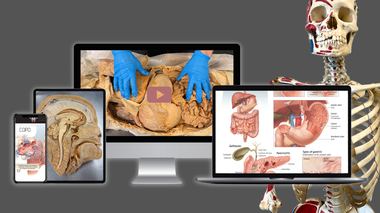
Introduction
The human musculoskeletal system is a masterpiece of biological engineering, providing structure, enabling movement, and safeguarding vital organs. Composed of bones, muscles, joints, tendons, and ligaments, this system exemplifies the intricate interplay of form and function in the human body. Anatomy as a whole, with its diverse systems and interconnected processes, remains foundational to understanding the marvel of human physiology.
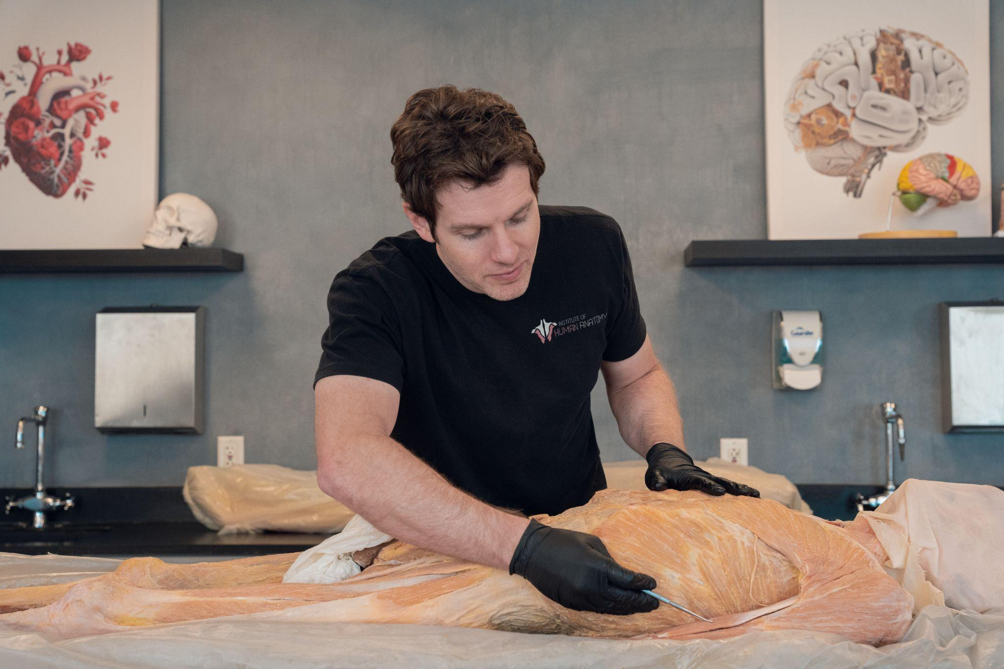
While textbooks and illustrations offer valuable insights true mastery demands hands-on human anatomy education. At the Institute of Human Anatomy (IOHA), we champion experiential learning through cadaver labs with online anatomical education that bridge theory and practice.
In this blog, we explore why tactile engagement with real cadavers is irreplaceable for mastering the musculoskeletal system and how our digital courses empower learners to excel.
The Musculoskeletal System: A Foundation for Movement and Health
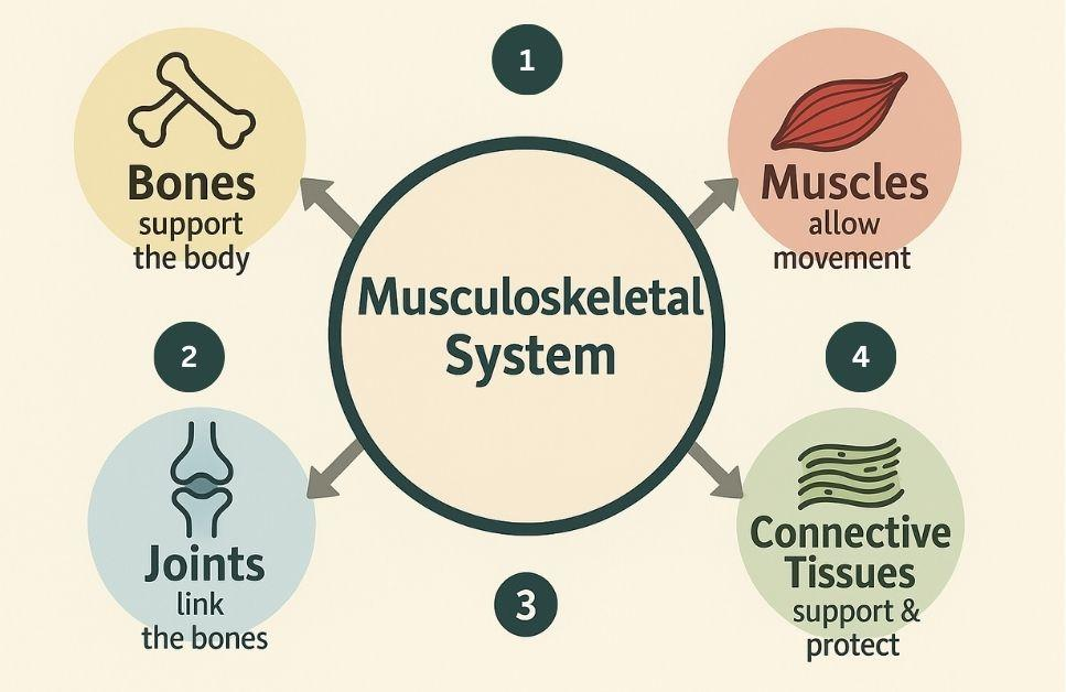
The musculoskeletal system’s primary roles, support, protection, and mobility are made possible by its intricate components:
- Bones: Serve as levers for movement, mineral reservoirs (storing calcium and phosphate), and factories for blood cell production.
- Joints: Facilitate motion with the help of synovial fluid, cartilage, and bursae.
- Connective Tissues: Link muscles to bones via tendons while ligaments stabilize joints.
- Muscles: Generate force via fascicle arrangements and tendon connections.
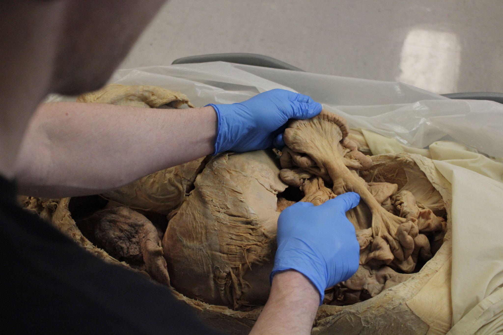
But grasping the complexity of these structures requires more than diagrams. For example, while radiography or MRI can detect bone remodeling or arthritis, only cadaveric study allows you to truly understand *the process, probing osteoblast-built trabeculae with your instruments, feeling the gritty erosion of arthritic cartilage, and witnessing how inflammation ravages synovial tissue in 3D space. Imaging shows* what happens; cadaver dissection reveals why and how, equipping you to interpret scans like a clinician, not just read them. This is where human dissection benefits shine.
Why Hands-On Human Anatomy Education Transcends Traditional Learning
1. Real-World Context for Theoretical Knowledge
Studying a human anatomy model can illustrate bone shapes or muscle origins, but cadavers reveal nuances textbooks can’t capture colon
- Bone Density Variations: the brittle texture of osteoporosis vs. the resilience of healthy bone. Learn more in our guide: Exploring the Skeletal System.
- Joint Mechanics: How synovial fluid reduces friction in a real knee joint.
- Muscle Fascicles: The multidirectional alignment of fibers dictates strength and movement of fibers dictating strength and movement.
At IOHA, learners gain access to high-definition cadaver-based videos that showcase real human anatomy, allowing them to observe tissue texture, identify pathologies, and see how lifestyle factors like calcium deficiency physically manifest in the body.
2. Developing Critical Clinical Skills
Active learning outcomes extend beyond memorization. Dissection hones
- Spatial Awareness: Tracing nerves or blood vessels through muscle layers.
- Surgical Precision: Handling delicate tissues without damaging adjacent structures.
- Diagnostic Insight: Identifying arthritis, tendinitis, or ligament tears firsthand.
These skills are vital for medical professionals, physical therapists, and fitness experts, all of whom benefit from anatomy courses grounded in hands-on, tactile learning.
3. Bridging the Gap Between Models and Reality
While anatomy mannequins are useful for basics, they lack the variability of human anatomy.
- Anomalies: Cadavers reveal natural variations (e.g., extra muscles or atypical nerve paths).
- Pathologies: Spotting bone spurs or cartilage degeneration in osteoarthritis.
- Texture and Elasticity: Feeling tendons’ toughness vs. ligaments’ flexibility.
As one IOHA student noted: “Seeing a model’s perfect femur is one thing, holding a real femur with osteoporotic pores changes how you think about patient care.”
Advantages of IOHA’s Cadaver Labs
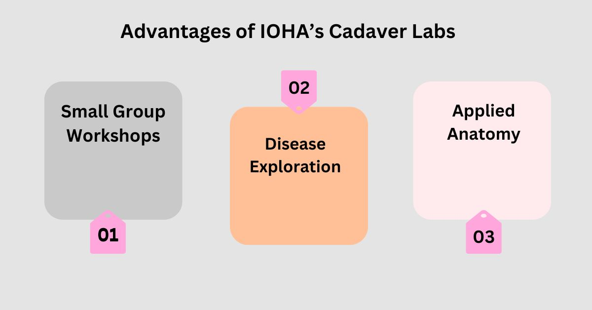
At the Institute of Human Anatomy, our hands-on human anatomy education programs are designed to maximize learning through:
- Small Group Workshops: Personalized guidance from expert instructors.
- Disease Exploration: Compare healthy vs. diseased tissues Such as rheumatoid vs. osteoarthritis).
- Applied Anatomy: Relate structures to clinical scenarios (e.g., rotator cuff injuries, ACL tears).
Our online courses are powered by high-fidelity 3D cadaver scans and live virtual dissections, allowing you to explore intricate anatomical variations and pathologies firsthand, no lab coat required. While models reinforce idealized structures, our cadaver-driven curriculum immerses you in the complexity of human tissues, preparing you to diagnose and treat with confidence
Diagnose Disorders Like a Pro: Insights from Real Cadavers
Hands-on learning isn’t confined to a lab. Our platform uses interactive cadaver modules to teach you how to identify and understand common musculoskeletal conditions:
- Osteoporosis: Virtually “palpate” cadaver bones to compare healthy vs. osteoporotic density, and see how fractures propagate in weakened structures. Resource: NIH Osteoporosis Guide.
- Arthritis: Manipulate 360° cadaver joint scans to spot cartilage erosion in osteoarthritis or synovial inflammation in rheumatoid Arthritis Foundation.
- Muscle Injuries: Zoom into torn fascicles or inflamed tendons in our cadaver image bank, paired with surgeon-led video analyses Muscle Health Myths .
Beyond Cadavers: The Role of Technology in Anatomy Education
Compare synovial joint degradation in osteoarthritis to inflammatory damage in rheumatoid arthritis.
- 3D Anatomy Mannequins: Reinforce spatial relationships (e.g., muscle attachments).
- Digital Scans: Complement dissection with CT/MRI correlations.
IOHA’s Cadaver-First Online Curriculum
Dive into anatomy with real cadaver scans and live virtual dissections, precision tools for mastering human variability. Our courses merge tactile skill-building with digital innovation:
Conclusion: Elevate Your Expertise with Hands-On Learning
Mastering the musculoskeletal system requires more than passive study. It demands immersion in its real-world complexity. Through IOHA workshops and cadaver labs, learners gain irreplaceable skills, from palpating bone textures to diagnosing joint disorders. Whether you’re a medical student, therapist, or fitness professional, hands-on human anatomy education transforms theoretical knowledge into actionable wisdom.
Ready to deepen your understanding? Explore our anatomy courses or contact us with any questions. The path to anatomical mastery starts here.

