
How Cadaver Videos Improve Anatomy Understanding
Dec 26, 2025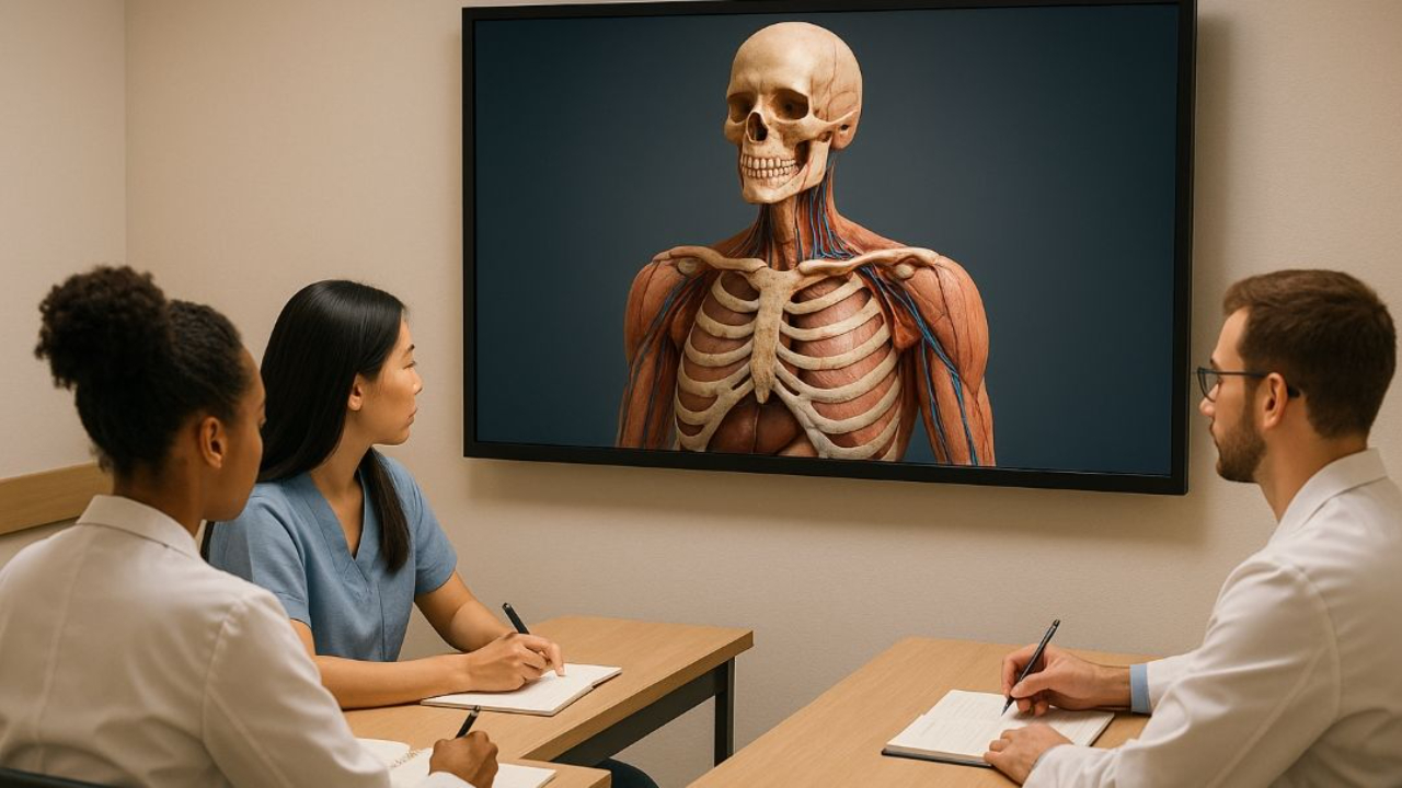
Cadaver videos are changing how anatomy is taught. These videos use real human cadavers to provide detailed, multi-angle views of the human body, helping students understand complex structures better than textbooks or static images. They’re widely accessible online, with platforms like the Institute of Human Anatomy reaching millions globally.
Key Benefits:
- 3D Visualization: Videos show spatial relationships between body parts, improving comprehension.
- Memory Retention: Students can replay content, boosting exam performance.
- Self-Paced Learning: Flexible study options reduce stress and suit diverse learning styles.
- Cost and Access: Videos address challenges like donor shortages, high costs, and limited lab access.
Studies confirm that combining cadaver videos with hands-on dissection leads to better learning outcomes, making them an essential tool in modern anatomy education.
How Cadaver Videos Improve Anatomy Learning
Better Visualization of 3D Structures
Grasping human anatomy isn't just about memorizing labels or locations - it's about understanding how body parts fit together in three-dimensional space. This is where cadaver videos shine, offering a unique advantage over traditional textbooks.
While textbooks rely on static images that force students to mentally piece things together, cadaver videos provide dynamic, multi-angle views of anatomical structures. This dynamic approach allows learners to see how different parts of the body connect and interact in ways that flat, two-dimensional images simply can't replicate. For example, these videos can clearly demonstrate the intricate relationships between blood vessels, nerves, and fascial planes, making complex spatial arrangements easier to understand. Research even shows that such videos help students form visual mental images, a critical skill for mastering anatomy through comprehension, visualization, and memorization.
Though virtual dissection platforms also contribute to spatial understanding, they lack the tactile experience of hands-on learning. Cadaver videos fill this gap, bridging theoretical knowledge with practical application. This enhanced visual clarity not only improves understanding but also boosts retention and performance in exams.
Improved Memory Retention and Exam Scores
Beyond improving understanding, cadaver videos have been shown to significantly enhance memory retention and exam results. Studies reveal that 89% of male students and 85% of female students found these videos helpful for memorization. In fact, virtual anatomy programs that incorporate videos often produce exam scores comparable to - or even better than - those achieved through traditional dissection methods. For instance, in neuroanatomy labs, students using virtual resources achieved higher average quiz scores than those relying solely on donor dissections.
Additionally, over 82% of male students and 83.7% of female students reported a deeper understanding of anatomy thanks to video resources, with about 90% of participants recommending them as an effective learning tool. The ability to replay and review complex structures makes videos particularly effective for reinforcing memory, especially when compared to text-based methods. They also cater to various learning preferences, offering a versatile tool for students with different study habits.
Self-Paced Learning for Different Learning Styles
Students learn in diverse ways, and cadaver videos cater to this diversity by offering flexible, self-paced learning opportunities. Research highlights that many students prefer dissection videos as part of their study routine, showcasing the importance of varied learning formats.
The self-paced nature of these videos allows students to pause, rewind, and rewatch as needed, making them especially useful for those who need extra time to grasp spatial relationships. This format also helps reduce pre-dissection anxiety. Watching real-life dissections in advance has been shown to significantly lower stress levels, making students more confident and prepared for hands-on sessions.
To enhance this flexibility, tools like the Institute of Human Anatomy’s AI Jonathan offer real-time, 24/7 support for anatomy and physiology questions. This personalized, on-demand feature helps students clarify concepts while watching videos, creating a more interactive and engaging learning experience. When combined with textbooks, digital guides, and other resources, cadaver videos become an integral part of a comprehensive, multimodal approach to mastering anatomy.
From Grant’s Dissection Video Collection: Reflection of the Abdominal Wall
Types of Cadaver Videos and Their Uses
Cadaver videos come in various formats, each tailored to specific educational goals. Knowing these differences helps students and educators select the most effective tools for their learning needs.
Complete Dissection Walkthroughs
Full-length dissection videos take viewers through entire anatomical regions, showing how structures connect and interact as the dissection progresses. These videos reveal sequential processes, system relationships, and spatial arrangements, helping students build a mental 3D map of the body. This approach not only aids understanding but also strengthens long-term memory.
One of the biggest advantages of complete walkthroughs is the context they provide. Watching an entire dissection allows students to see how blood vessels branch, nerves weave through muscles, and fascial planes divide compartments - details that shorter videos might skip.
Studies back up the effectiveness of these videos. Research has shown that virtual and hybrid learning models can achieve exam results on par with traditional in-person methods, with no evidence of in-person instruction consistently outperforming these alternatives. Additionally, full dissection videos are excellent prep tools for lab sessions, helping students enter with clearer expectations and a solid foundation for hands-on learning.
Region-Specific and System-Specific Tutorials
For those who need to focus on particular areas, region- or system-specific videos are invaluable. These tutorials zero in on specific parts of the body, like the cardiovascular system, musculoskeletal structures, or neuroanatomy, making them ideal for exam prep or targeted review.
Unlike full-length dissections, these focused videos save time by skipping unrelated material. For example, a student studying for a cardiovascular exam can repeatedly review the heart and major vessels without wading through other systems. This approach works well with anatomy courses that break content into smaller, manageable units.
Neuroanatomy, in particular, benefits from this targeted format. Research has shown that students using virtual neuroanatomy videos scored higher on quizzes and reported better understanding, spatial ability, and satisfaction compared to those relying solely on donor dissections. Additionally, region-specific videos cater to diverse learning styles. For instance, a study found that 46.5% of male and 41.8% of female students preferred PowerPoint presentations, while 32.8% of male and 36.8% of female students specifically chose cadaver dissection videos.
Interactive and Student-Created Videos
Interactive videos and student-produced content are gaining traction as engaging alternatives in anatomy education. Interactive features, like embedded questions or adjustable viewing angles, encourage active participation, which can boost comprehension and retention.
Student-created videos add another layer of relatability. When advanced students produce educational content, their explanations often reflect their own learning experiences, making the material easier for peers to grasp. Plus, creating these videos reinforces the creators’ knowledge while benefiting others.
One standout example is the Institute of Human Anatomy’s AI tool, Jonathan, which offers real-time, 24/7 support for anatomy and physiology questions. This AI assistant provides instant answers, detailed explorations of major body systems, and personalized study help, all while students engage with video content.
Interactive and peer-created videos also address common challenges in virtual anatomy learning. Studies show that students learning anatomy virtually in isolation often feel less engaged compared to those using hybrid methods. By incorporating active learning and peer perspectives, these video formats create a more engaging, hands-on experience that promotes deeper understanding and better retention.
Using Cadaver Videos Alongside Hands-On Dissection
Cadaver videos shine when they complement, rather than replace, hands-on dissection. While videos provide excellent visual learning opportunities, they cannot replicate the physical experience of working with real tissues. By combining both methods, educators can create a well-rounded and effective anatomy curriculum.
Videos and Dissection: A Perfect Pairing
Hands-on cadaver dissection offers something unique: the tactile experience of feeling tissue textures, navigating fascial planes, and developing spatial awareness - skills critical for clinical practice. On the other hand, videos bring multi-angle views, highlight anatomical variations, and allow repeated reviews of intricate relationships. Together, these methods teach classification, spatial relationships, and three-dimensional perspectives, while also building tactile skills and fostering a deeper understanding of human anatomy.
Videos also serve as excellent preparatory tools, helping students feel more confident and understand what to expect in the lab. During dissections, they provide alternative perspectives and step-by-step guidance. Afterward, they reinforce learning and assist with exam preparation.
Research shows that students are more engaged when hands-on dissection is paired with virtual tools. In contrast, students relying solely on virtual anatomy courses often disengage at higher rates compared to those in hybrid programs.
Research on Blended Learning Approaches
Studies consistently support the combination of videos with physical dissection. Blended approaches that integrate cadaver labs with video training have proven to be both cost-efficient and highly effective for students. In fact, research shows that hybrid models can produce exam results that rival - or even surpass - those of traditional, in-person anatomy courses. Students overwhelmingly report that video content helps them better memorize and recall anatomical details.
This approach also benefits surgical residents. Supplementing cadaveric dissection with videos boosts confidence, deepens anatomical knowledge, and sharpens surgical skills. The most effective programs combine cadaver labs with textbooks, multimedia tools, and videos, avoiding reliance on a single teaching method.
To ensure quality, expert educators should be involved in creating or reviewing cadaver videos. This guarantees that the materials are accurate and effectively enhance the hands-on dissection experience.
A blended approach doesn’t just improve learning outcomes - it also addresses practical challenges like donor shortages, high costs, ethical concerns, and disruptions caused by events like pandemics. By integrating high-quality video content with cadaver resources, institutions can make anatomy education more accessible without compromising on quality.
The Institute of Human Anatomy showcases this strategy, combining interactive cadaver videos with hands-on dissection to deliver a modern, comprehensive anatomy education experience.
Access and Quality Standards for Cadaver Videos
Expanding Access to Anatomy Education
Traditional cadaver dissection labs come with significant challenges, limiting access to quality anatomy education for many students. Geographic barriers often prevent hands-on learning opportunities, but cadaver videos are changing the game by making anatomy education available to anyone with an internet connection.
The numbers speak for themselves: 93.2% of male and 89.3% of female medical students use YouTube as a resource for anatomy learning. This widespread use highlights the way videos overcome obstacles such as limited lab hours, restricted access to cadaver facilities, and the hefty costs tied to body donor programs.
What’s more, videos let students revisit material at their own pace - pausing, rewinding, and reviewing complex structures as needed. This flexibility is a lifesaver for students in remote areas, those attending institutions without cadaver labs, or anyone juggling demanding medical school schedules.
The reach of cadaver video content is staggering. For example, the Institute of Human Anatomy has amassed over 2 billion video views and boasts a following of more than 24 million subscribers as of December 2025. Such platforms are breaking down location and resource barriers, bringing anatomy education to a global audience.
Cost is another factor driving the shift to video-based learning. Running cadaver labs involves steep expenses for donor procurement, facility upkeep, and managing chemicals like formaldehyde. Videos eliminate these costs while offering scalable education for countless students over time. For schools with tight budgets, high-quality video content provides an affordable alternative without sacrificing educational impact.
Safety is also a concern. Traditional dissection labs expose students to chemicals like formaldehyde, which can lead to respiratory issues or allergic reactions. Videos eliminate these risks entirely. Plus, watching dissections on video before participating in person can ease anxiety for students.
Platforms like the Institute of Human Anatomy are taking accessibility even further with tools like AI Jonathan, a 24/7 virtual guide that provides real-time answers and personalized study support. This kind of on-demand assistance addresses time constraints and offers individualized help that traditional labs can’t match.
As cadaver videos become more widely available, ensuring their quality is critical for effective learning.
How to Evaluate Video Quality and Educational Content
With the growing availability of cadaver videos, maintaining high standards is essential. Not all videos are created equal, so students and educators need clear criteria to identify the best resources. Key factors include expert involvement, visual clarity, and educational value.
First, expert oversight is non-negotiable. Videos should be created or reviewed by qualified anatomy educators to ensure accuracy and alignment with medical curricula. Look for creators who list their credentials to verify their expertise.
Next, visual quality matters. Effective videos should provide clear, well-lit views of anatomical structures from multiple angles. Good camera work helps illustrate the three-dimensional relationships between organs, tissues, and vessels - something static images just can’t achieve. Audio narration should explain these structures in clear, accessible language, using proper anatomical terms.
Evidence of educational effectiveness is another important measure. Research shows that virtual and hybrid learning models incorporating videos often outperform traditional in-person instruction. For example, in neuroanatomy studies, students using virtual dissection scored significantly higher on quizzes than those relying solely on donor dissections (p < 0.05). These findings highlight how well-designed videos can match - or even surpass - traditional methods.
Student satisfaction also offers valuable insights. Surveys reveal that 90.4% of male and 87.3% of female students recommend YouTube for anatomy learning. Positive feedback on understanding and retention is a strong indicator of a video’s quality.
Variety in content formats is crucial to accommodate different learning styles. While some students prefer PowerPoint presentations (46.5% of males and 41.8% of females), others lean toward cadaver dissection videos (32.8% of males and 36.8% of females). High-quality video libraries should offer diverse options, including full dissection walkthroughs, region-specific tutorials, system-focused content, and presentation-style materials. This ensures resources are accessible to visual, auditory, and structured learners alike.
Curriculum alignment is another critical factor. Videos should support specific learning objectives and integrate seamlessly with anatomy courses. The best content goes beyond identifying structures to explore anatomical variations, clinical applications, and spatial relationships, helping students connect theory to practice.
Accessibility features also play a big role. Videos should work across various devices and internet speeds, with offline viewing options whenever possible. Closed captions and transcripts are essential for students with hearing impairments or those learning in non-native languages, broadening the audience who can benefit from the content.
Institutions should establish review processes to evaluate video resources. Accuracy, clarity, and educational value should all be assessed, with input from both experts and students. Feedback surveys and performance metrics can help confirm whether videos are improving learning outcomes.
The Institute of Human Anatomy sets a strong example by combining expert-created cadaver videos with digital study guides and interactive tools. By prioritizing high production standards and involving qualified anatomists, platforms like this ensure that expanded access doesn’t come at the expense of educational quality.
Conclusion: The Future of Cadaver Videos in Anatomy Education
Cadaver videos have become an invaluable resource in teaching anatomy, delivering results that often rival traditional dissection methods. Studies reveal that these videos not only help students grasp and retain complex concepts but, in some cases, virtual learning even outperforms hands-on approaches in exams. By addressing challenges like geographic limitations, restricted lab access, high costs, and concerns about formaldehyde exposure, cadaver videos are reshaping how anatomy is taught.
Combining cadaver videos with other resources - like physical dissections, textbooks, and interactive tools - offers students a well-rounded learning experience. The Institute of Human Anatomy is leading this transformation, blending expertly crafted cadaver videos with cutting-edge technology. Their introduction of AI Jonathan, a virtual guide available 24/7, marks a leap forward by providing personalized, real-time study support. This approach highlights how artificial intelligence can complement expertly produced cadaver content, making education more accessible and tailored to individual needs.
The future of anatomy education lies in further integrating AI with cadaver videos. Students can revisit intricate anatomical structures as often as needed, receive expert guidance on demand, and enjoy a learning experience tailored to their specific goals. Rather than replacing traditional dissection, these advancements enhance and expand its potential.
As universities face reduced lab hours, donor shortages, and other logistical hurdles, high-quality cadaver videos are set to play an even greater role in anatomy programs. By ensuring these videos maintain expert oversight, clear visuals, and alignment with educational goals, they open the door to comprehensive training for anyone with internet access. This shift not only addresses current challenges but also redefines how anatomy education can reach students around the world.
FAQs
How do cadaver videos help students better understand anatomy compared to traditional textbooks?
Cadaver videos offer an immersive and visually rich method to study human anatomy, bringing a level of detail and authenticity that textbooks simply can't match. By showcasing real human cadavers, these videos reveal the intricate structures of muscles, organs, and tissues in their natural context, helping to connect theoretical knowledge with real-world application.
Unlike the fixed images in textbooks, cadaver videos illustrate how anatomical structures relate to one another spatially and functionally within the body. This dynamic, visual approach simplifies complex concepts and enhances understanding, making it a valuable resource for students, educators, and healthcare professionals.
How do cadaver videos enhance learning when paired with hands-on dissection?
Cadaver videos serve as a powerful visual tool, enhancing the hands-on experience of dissection by clearly illustrating anatomical structures and their connections. They give students the flexibility to review intricate concepts at their own pace, helping to solidify what they’ve learned during lab sessions.
When paired with dissection, these videos offer a blend of theoretical insight and practical application. This combination not only deepens students’ understanding of human anatomy but also boosts their ability to retain information and confidently apply their knowledge in practical settings.
How do cadaver videos help make anatomy education more affordable and accessible?
Cadaver videos offer an affordable and convenient way to study human anatomy through detailed visual demonstrations of actual human bodies. They remove the barriers of needing physical access to cadaver labs, which are often costly and not available everywhere.
With cadaver videos, learners - whether medical students or healthcare professionals - can study whenever and wherever they want. These videos also allow users to revisit challenging topics as often as needed, making anatomy education more accessible and practical for a diverse group of learners.

