
Unraveling the Neural Network: A Deep Dive into the Human Nervous System Through Dissection
Jun 18, 2026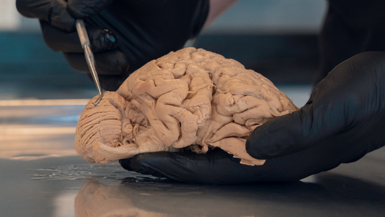
The human body is an intricate marvel, a symphony of interconnected systems working in harmony to sustain life. At the heart of this biological masterpiece lies the human nervous system, a complex, high-speed communication network that orchestrates every thought, movement, sensation, and emotion. It is the command center, the interpreter, and the responder, allowing us to interact with the world and understand ourselves.
For anyone pursuing anatomy education, whether students, medical professionals, therapists, or estheticians, truly grasping the intricacies of this neural network is paramount. While textbooks, digital models, and lectures provide foundational knowledge, many educators consider cadaveric dissection one of the most powerful methods for gaining deep insight into its architecture. This hands-on approach transcends theoretical understanding, revealing the three-dimensional reality and delicate relationships of the nerves, brain, and spinal cord in a way no other medium can.
In this comprehensive exploration, we will embark on a journey through the neuroanatomy of the human nervous system. We will demystify its major components, delve into their functions, and underscore why the experience of cadaveric dissection is widely regarded as a central component of profound anatomical study of this vital system, particularly when combined with other modern teaching methods.
The Human Nervous System: An Architect's Blueprint of Life
Imagine a vast, sophisticated supercomputer connected to an equally vast sensor and effector network throughout your entire body. This is, in essence, your human nervous system. Its primary role is to collect, process, and respond to information, both from the external world and from within your body. It allows us to perceive a sunset, feel the warmth of a hug, learn a new skill, regulate our heartbeat, and even dream.
Functionally, the nervous system performs three core tasks:
- Sensory Input: Gathering information from internal and external stimuli (e.g., touch, sight, temperature, internal organ states).
- Integration: Processing and interpreting the sensory input, deciding what action, if any, needs to be taken. This is where higher cognitive functions, memory, and emotion reside.
- Motor Output: Executing responses to the integrated information by activating muscles or glands.
Structurally, the human nervous system is traditionally divided into two main parts:
1. The Central Nervous System (CNS)
The CNS is the ultimate control center, comprising the brain anatomy and the spinal cord. It is protected by bone (the skull and vertebral column) and cushioned by cerebrospinal fluid (CSF).
2. The Peripheral Nervous System (PNS)
The PNS consists of all the peripheral nerves that extend from the CNS to the rest of the body, including muscles, organs, and sensory receptors. It acts as the communication link between the CNS and the periphery.
Let's dive deeper into each of these incredible divisions.
Delving into the Central Nervous System (CNS)
The CNS is where the magic happens, where thoughts are formed, memories are stored, and every bodily function is coordinated.
The Brain: The Ultimate Command Center
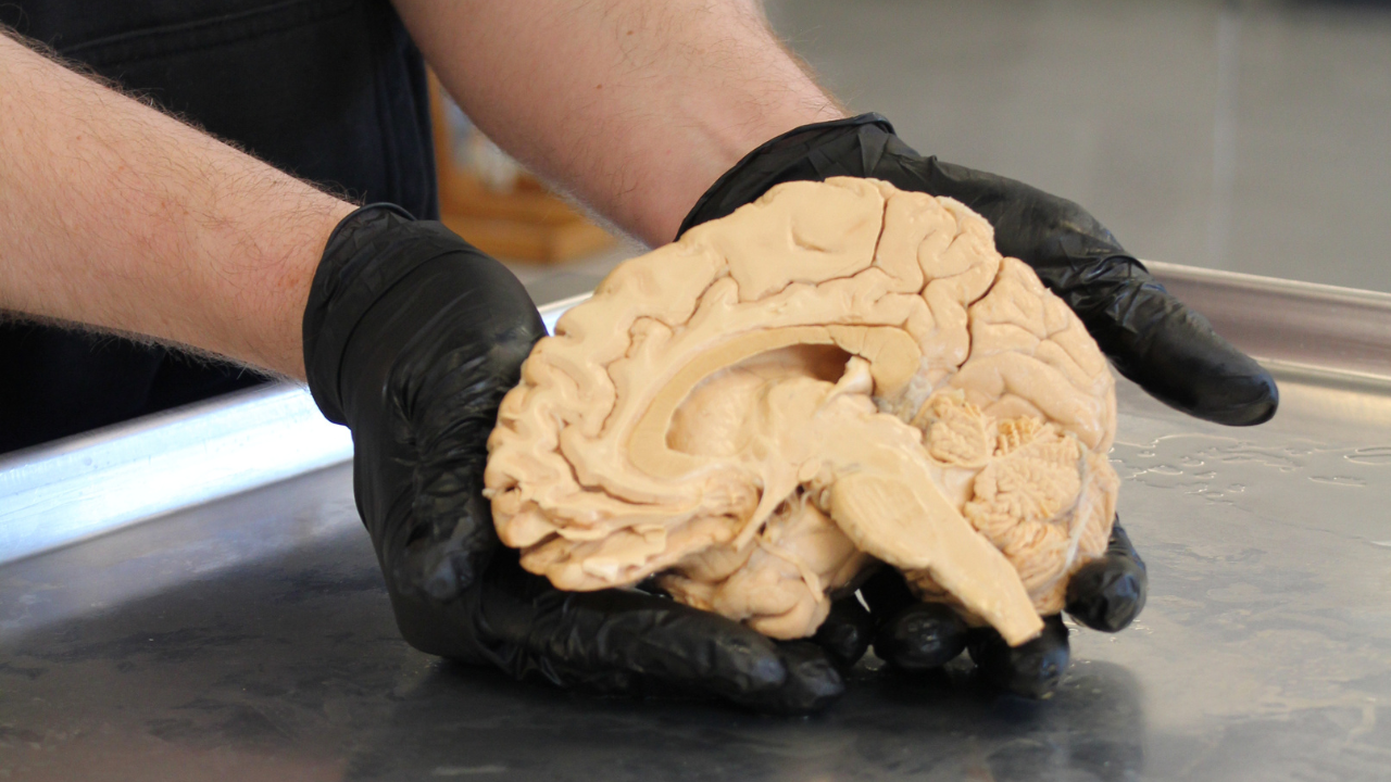
Weighing in at approximately three pounds, the human brain is is one of the most complex known biological structures. It's the epicenter of our consciousness, personality, and intellect. A deep dive into brain anatomy reveals fascinating structures, each contributing to our overall being.
Major Regions of the Brain:
- Cerebrum: The largest part of the brain, responsible for higher functions like thought, language, memory, and voluntary movement. Its surface, the cerebral cortex, is characterized by folds (gyri) and grooves (sulci) that dramatically increase its surface area, allowing a greatly expanded cortical surface area and dense packing of neurons. It is divided into four main lobes:
- Frontal Lobe: Involved in planning, decision-making, problem-solving, personality, and voluntary motor control.
- Parietal Lobe: Processes sensory information like touch, temperature, pain, and pressure, and integrates sensory data to create a spatial map of our body and environment.
- Temporal Lobe: Essential for processing auditory information, memory formation, and language comprehension.
- Occipital Lobe: Primarily responsible for processing visual information.
- Cerebellum: Located at the back of the brain, beneath the cerebrum. Often called the "little brain," it plays a crucial role in coordinating voluntary movements, balance, posture, and motor learning. Without it, our movements would be clumsy and uncoordinated.
- Brainstem: Connects the cerebrum and cerebellum to the spinal cord. It's a vital structure that controls many essential involuntary functions, including breathing, heart rate, blood pressure, sleep cycles, and consciousness. It comprises the midbrain, pons, and medulla oblongata.
- Diencephalon: Situated between the cerebrum and brainstem, it contains the thalamus (relay station for sensory information) and the hypothalamus (controls essential bodily functions like hunger, thirst, sleep, and body temperature, and links the nervous system to the endocrine system).
Understanding the intricate three-dimensional relationships between these structures is paramount for any profound anatomical study. Traditional 2D diagrams can only offer a limited perspective; it's within the context of a tangible specimen that the true marvel of brain anatomy unveils itself.
The Spinal Cord: The Information Superhighway
Extending from the brainstem down through the vertebral column, the spinal cord is a cylindrical bundle of nervous tissue that serves as the main conduit for information between the brain and the rest of the body. It's not just a passive pathway; it also houses neural circuits that mediate reflexes, allowing for rapid, involuntary responses to stimuli without direct input from the brain.
Structure of the Spinal Cord:
- Gray Matter: Located centrally, shaped like a butterfly, and composed primarily of neuron cell bodies, dendrites, and unmyelinated axons. This is where information processing occurs.
- White Matter: Surrounds the gray matter and consists of myelinated axons, organized into tracts (bundles of nerve fibers). These tracts carry sensory information up to the brain and motor commands down from the brain.
- Protection: The spinal cord is meticulously protected by three layers of meninges (dura mater, arachnoid mater, pia mater) and bathed in cerebrospinal fluid, which provides cushioning and nourishment.
Injuries to the spinal cord can have devastating consequences, underscoring its critical role in motor control, sensation, and autonomic functions. For medical students, therapists, and practitioners, a thorough anatomical study of the spinal cord is non-negotiable for understanding neurological conditions and designing effective treatments.
Exploring the Peripheral Nervous System (PNS)
While the CNS is the command center, the Peripheral Nervous System is the vast network of lines connecting it to every corner of your body. It's composed of all the peripheral nerves that branch out from the brain and spinal cord, conveying signals to and from the CNS.
Divisions of the PNS:
The PNS is often described in terms of structural (cranial and spinal nerves) and functional components, including the somatic, autonomic, and enteric systems:
- Somatic Nervous System (SNS): This system is responsible for voluntary control of skeletal muscles and carries sensory information from the body's sensory organs (like skin, muscles, and joints) back to the CNS. It's how you consciously move your arm or feel a gentle touch.
- Cranial Nerves: Twelve pairs of nerves emerge directly from the brain or brainstem. They control a wide range of functions, including smell, vision, eye movement, facial expressions, taste, hearing, balance, and aspects of internal organ control. For example, the optic nerve (cranial nerve II) transmits visual information, while the vagus nerve (cranial nerve X) contributes to regulation of heart rate and digestion
- Spinal Nerves: Thirty-one pairs of nerves that branch off the spinal cord at different levels, serving specific dermatomes (areas of skin) and myotomes (groups of muscles). These nerves often combine to form complex networks called plexuses (e.g., brachial plexus, lumbar plexus) that innervate the limbs.
- Autonomic Nervous System (ANS): This system regulates involuntary bodily functions that are not under conscious control, such as heart rate, digestion, respiration, blood pressure, and pupil diameter. It operates largely outside our conscious awareness to maintain homeostasis. The ANS is further divided into sympathetic, parasympathetic, and enteric divisions, with sympathetic and parasympathetic branches often having opposing effects in many organs:
- Sympathetic Nervous System: Often referred to as the "fight or flight" system, it prepares the body for stress or emergencies. It increases heart rate, dilates pupils, inhibiting some aspects of digestion, and redirecting blood flow to muscles.
- Parasympathetic Nervous System: Known as the "rest and digest" system, it promotes restorative and maintenance functions, such as slowing heart rate, constricting pupils, and stimulating digestion.
- Enteric Nervous System (ENS): A network of neurons in the walls of the gastrointestinal tract that can coordinate many aspects of gut function locally; it is frequently considered a third division of the autonomic or peripheral nervous system
The intricate pathways of peripheral nerves are complex. Damage to these nerves can lead to issues such as numbness, tingling, muscle weakness, or paralysis, reflecting the PNS role in motor control, sensation, and autonomic regulation. Understanding their precise origins, courses, and destinations is crucial for diagnosing and treating neurological conditions, making detailed anatomical study of the PNS indispensable.
Why Cadaveric Dissection is Indispensable for Neuroanatomy
In an era of advanced digital learning tools and virtual reality, one might question the continued relevance of cadaveric dissection. However, for fields requiring a truly profound understanding of the human nervous system, particularly neuroanatomy, the experience of working with human cadavers is widely regarded as uniquely valuable.
The Unrivaled Realism of Cadavers
While sophisticated digital models and simulations offer fantastic supplementary resources (and you can explore some of these through our digital resources), they cannot fully replicate the nuanced, three-dimensional complexity, texture, and individual variations found in a real human body.
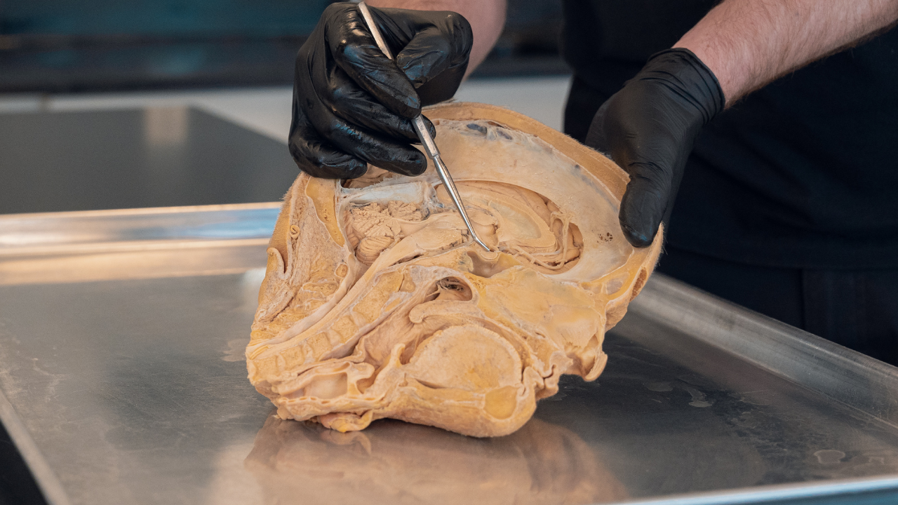
- Direct Observation of Anatomical Relationships: The nervous system, especially the brain anatomy and its connections to the spinal cord and peripheral nerves, is characterized by incredibly delicate structures nestled among other tissues. During cadaveric dissection, students meticulously peel back layers, observing how nerves weave through muscles, blood vessels, and fascia. This process reveals the true spatial relationships and topographical organization that static images or digital models can only approximate. You see the nerves emerging from the spinal cord, branching out to form plexuses, and then fanning out to their target tissues.
- Understanding Texture and Consistency: Neural tissue has a unique consistency—the gelatinous nature of the brain and the firm yet delicate texture of nerves. These tactile sensations are critical for future clinicians who will palpate structures, perform injections, or conduct surgical procedures. This haptic feedback is not reproduced by current digital tools.
- Appreciating Variability: No two human bodies are exactly alike. Cadaveric dissection exposes students to the natural anatomical variations that exist, preparing them for the diversity they will encounter in clinical practice. This understanding is particularly important for the human nervous system, where slight deviations can have significant implications.
- Demystifying Intricate Pathways: The pathways of nerve impulses are not always straightforward. Nerves twist, turn, and converge. Tracing these delicate structures through their entire course during cadaveric dissection is the most effective way to internalize these complex routes. It brings neuroanatomy to life, making abstract concepts concrete.
Inside the Brain of a Cadaver | Pathways in the brain
Enhancing Anatomy Education Through Hands-On Learning
The educational benefits of cadaveric dissection extend far beyond mere observation. It fosters a unique and deep learning experience that shapes competent and compassionate professionals.
- Active Learning and Critical Thinking: Dissection is an active process of discovery. It requires critical thinking, problem-solving, and decision-making as students identify structures, trace pathways, and correlate theoretical knowledge with practical findings. This process is invaluable for developing diagnostic understanding and surgical acumen.
- Developing Precision and Dexterity: Working with delicate neural structures demands meticulous attention to detail and fine motor skills. This hands-on experience sharpens a student's dexterity, a crucial skill for any medical or therapeutic profession. Our workshops, like those detailed in our Workshop FAQs, are designed to cultivate these essential skills.
- Reinforcing Theoretical Knowledge: The tactile experience of identifying a cranial nerve or tracing a specific tract within the spinal cord deeply ingrains the theoretical knowledge learned from textbooks. It transforms abstract facts into tangible realities.
- Fostering Respect and Professionalism: Working with human cadavers instills a profound sense of respect for the human body and life itself. It's a privilege to learn from those who have donated their bodies to science, and this experience often cultivates empathy and professionalism in students, shaping them into more compassionate practitioners. You can learn more about our philosophy and mission on our ABOUT page.
The Process of Anatomical Study: From Theory to Practice
A comprehensive anatomical study of the human nervous system involves a multi-faceted approach. It begins with theoretical learning, lectures, textbooks, and interactive models. However, it truly comes alive in the anatomy lab.
Students of neuroanatomy embark on a journey that combines classroom instruction with practical, hands-on experience. They learn about the cellular components of neurons, the chemistry of nerve impulses, and the functions of different brain regions. Then, they enter the dissection lab, where they apply this knowledge directly.
Using specialized Anatomy Tools, students carefully remove layers of tissue, identify landmarks, and meticulously expose the delicate structures of the human nervous system. This is where the intricacies of brain anatomy, the branching patterns of the spinal cord nerves, and the extensive network of peripheral nerves are unveiled.
This hands-on experience is critical not only for future surgeons and physicians but also for massage therapists, bodyworkers (explore our Massage Therapy & Bodywork In-Lab Workshop), and estheticians (learn more about Facial Anatomy & Facial Esthetics) who need a precise understanding of nerve pathways to safely and effectively perform their work.
The precision and meticulousness required for cadaveric dissection train individuals to be detail-oriented, patient, and systematic—qualities essential for any profession that interacts directly with the human body. The opportunity to directly visualize the impact of structures on function helps cement understanding in a way that passive learning cannot achieve.
Challenges and Rewards of Neuroanatomy Dissection
Dissecting the human nervous system is not without its challenges, but many students and educators judge the educational rewards to be substantial.
Challenges
- Delicate Nature of Neural Tissue: Brain and nerve tissues are notoriously delicate and require extreme care to avoid damage during dissection. This teaches students patience and fine motor control.
- Complexity of Pathways: The sheer number of interconnections and the often-microscopic scale of some neural pathways can be daunting. Tracing these networks requires keen observation and a strong foundational knowledge.
- Time Commitment: Thorough cadaveric dissection of the nervous system is time-consuming and labor-intensive, often requiring many hours in the lab.
- Emotional Aspect: Working with human cadavers can be emotionally challenging, requiring students to develop coping mechanisms and maintain a respectful, professional demeanor.
Rewards
- Profound Understanding: The most significant reward is a deep, three-dimensional understanding of the human nervous system that is difficult to match with other single methods alone.
- Enhanced Clinical Skills: The practical experience is perceived to contribute to improved diagnostic capabilities, better surgical planning, and more effective patient care.
- Problem-Solving Skills: Identifying structures, understanding their relationships, and navigating anatomical variations cultivates critical thinking and problem-solving abilities.
- Deep Appreciation for Life: Experiencing the intricate design and vulnerability of the human body through dissection fosters a deep appreciation for life and the responsibility that comes with caring for others. It often sparks what we call "aha!" moments, where complex theoretical concepts suddenly click into place with tangible reality.
The Future of Anatomy Education
The landscape of anatomy education is continually evolving. We are seeing a powerful integration of traditional methods, like cadaveric dissection, with cutting-edge technology. Tools like virtual reality, augmented reality, and AI-powered learning platforms (such as our own AI JONATHAN) are becoming increasingly prevalent, offering new ways to visualize and interact with anatomical structures.
However, these technologies are best utilized as supplements, not replacements, for the hands-on experience of cadaveric dissection.
Blended learning approaches, combining the best of both worlds, are increasingly recommended as a highly effective strategy for a comprehensive anatomical study.
The tactile, observational, and problem-solving skills honed through dissection are widely regarded as central to developing the practical competence and professional judgment essential for medical and healthcare practitioners.
For those looking to deepen their understanding, whether through a foundational course or specialized workshops, exploring available resources is key. You can find a variety of options, from Anatomy Study Bundles to All Courses, designed to enhance your anatomy education. Continuing professional development is vital, and the insights gained from direct cadaveric dissection are enduring.
Conclusion
The human nervous system stands as the ultimate testament to biological engineering—a complex, dynamic network that defines our very existence. Its intricate design, from the vast expanses of brain anatomy to the delicate pathways of the spinal cord and the far-reaching branches of peripheral nerves, demands a profound level of understanding for anyone dedicated to health and wellness.
While technological advancements offer incredible avenues for learning, the role of cadaveric dissection in neuroanatomy remains paramount. It offers a uniquely valuable, hands-on opportunity to truly unravel the neural network, providing insights into spatial relationships, textures, and variations that current alternative methods do not fully replicate. It demystifies complexity, builds essential skills, and instills a deep respect for the human body, shaping proficient, empathetic professionals.
By embracing the unique opportunities provided by cadaveric dissection, students and professionals alike can gain a foundational understanding that is difficult to match by other single methods of the human nervous system, empowering them to contribute more effectively to the well-being of others.
The journey into the human nervous system through dissection is not just an anatomical study; it's a deep dive into the essence of what makes us human. We encourage you to explore our Blog Articles for more insights and resources on this incredible journey.

