
How Cadavers Improve Regional Anatomy Learning
Jan 28, 2026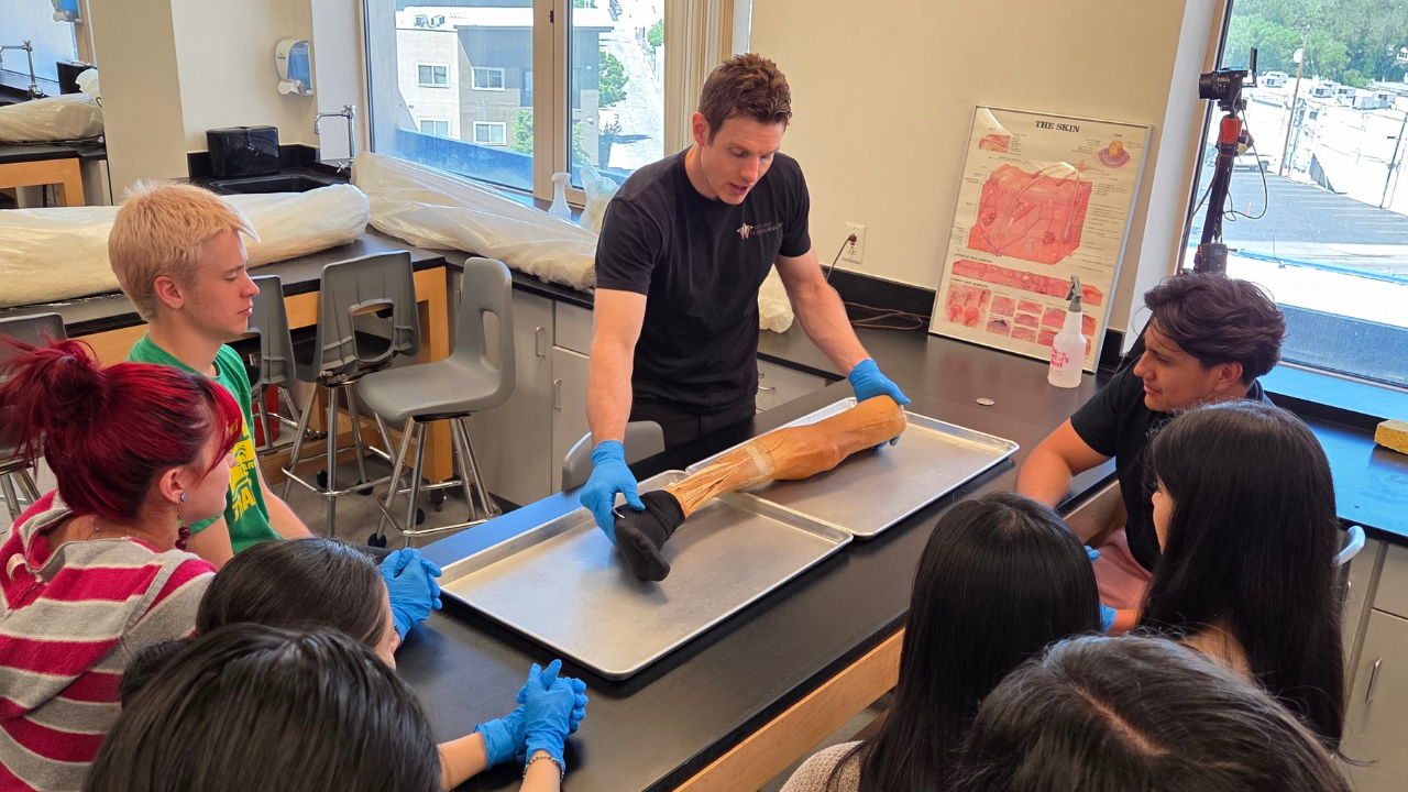
Cadaver-based learning is one of the most effective ways for medical students to understand regional anatomy. Unlike digital tools or plastic models, cadavers provide hands-on experience with real human tissue, helping students grasp spatial relationships, anatomical variations, and tactile feedback. Studies show that:
- Students trained with cadavers perform better on tests: For example, dissection groups scored significantly higher than those using models or software.
- Confidence improves: 64% of cadaver-trained students feel confident understanding trauma, compared to 15.4% of virtual-only learners.
- Clinical skills are enhanced: Cadaver training sharpens motor skills, spatial reasoning, and the ability to interpret imaging like X-rays and CT scans.
- Emotional learning matters: Many students develop empathy and respect for the human body through cadaver work, which translates into professionalism in their careers.
While modern tools like 3D software offer value, cadavers remain unmatched in preparing students for complex medical procedures. They provide a realistic, tactile, and emotionally engaging way to learn anatomy that directly impacts patient outcomes.
What Research Shows About Cadaver-Based Learning
Better Knowledge Retention and Understanding
Research strongly supports the benefits of cadaver-based learning for mastering anatomy. For instance, a study conducted between 2014 and 2019 at the University of Thessaly in Greece examined 313 first-year medical students learning upper limb anatomy. These students were divided into four groups: dissection, prosections, plastic models, and 3D software. The group that practiced dissection scored significantly higher on comprehension tests compared to the others.
"Dissection remains first in students' preferences and achieves higher knowledge acquisition." – Scientific Reports
But the advantages go beyond just test scores. A 2015 study from the University of Heidelberg in Germany compared 238 students during their first-year gross anatomy exams. Students who used cadaver-specific CT scans alongside traditional dissection performed 27% better, scoring an average of 21.8, than those in the conventional anatomy group. Additionally, students overwhelmingly rate human tissue as the most effective tool for learning outcomes such as 3D visualization, identifying anatomical variations, and understanding functional anatomy. Notably, 95% of medical students and 98% of dental students found cadaver dissection to be highly beneficial. To maximize these benefits, students can also apply evidence-based study strategies, as outlined in How to Study Anatomy: 5 Proven Memory Techniques.
The BEST Way to Learn ANYTHING (Especially Anatomy)!!! | Institute of Human Anatomy
Increased Confidence and Practical Skills
Hands-on cadaver training not only sharpens practical skills but also builds confidence, a critical factor in clinical decision-making. A study conducted at Rowan University School of Osteopathic Medicine between 2020 and 2021 compared two groups of medical students: one trained in-person with cadavers and the other using virtual labs during the COVID-19 pandemic. The results were striking. Among cadaver-trained students:
- 64% reported high confidence in understanding trauma, compared to just 15.4% of virtual-only students.
- 72% felt comfortable interpreting X-rays and CT scans, versus 30.9% in the virtual group.
- 90% were confident identifying pathology in radiographic images, compared to 65.4%.
- Proficiency in point-of-care ultrasound skills was 64% for cadaver-trained students, while only 36.9% of virtual-only students felt the same.
"The process of dissecting tissue provides an appreciation for location and layers of tissues that are less clear with the plastinated models." – Dental Resident, University of Texas Health Science Center at Houston
Tactile and Emotional Learning Benefits
One of the unique aspects of cadaver dissection is the tactile experience it offers, something digital tools simply cannot replicate. Students can physically touch and manipulate structures, like nerves and arteries, to understand their textures and spatial relationships. In fact, 63% of students found this hands-on approach particularly valuable for deepening their knowledge of anatomical variations.
Dissection also fosters emotional learning. Many students view their cadaver as their "first patient", which helps cultivate empathy and respect - qualities essential for medical practice. In one study, all participants expressed a deep sense of gratitude and respect for their donors. Furthermore, 88% of students who trained with cadavers reported an increased respect for the human body, compared to just 33.3% of those who trained virtually. This emotional connection often translates into greater professionalism and resilience in clinical settings.
"Many students demonstrated a shift in thinking about their cadaver as less of a tool and more of a patient, which allowed them to generalize their experience in lab towards their future careers in medicine." – Erin Parker, Uniformed Services University of the Health Sciences
These findings underline how cadaver-based learning plays a crucial role in developing both technical and emotional skills, setting the stage for improved clinical expertise - an area explored further in the next section.
What It's Like Working With Dead Bodies
Why Cadavers Work Better Than Other Methods
Cadaver vs Digital Learning Methods: Effectiveness Comparison for Medical Students
When it comes to learning anatomy, cadavers provide an advantage that digital tools and plastic models simply can't replicate. While these alternatives have their place in education, they fall short of delivering the depth and hands-on understanding that real human tissue offers.
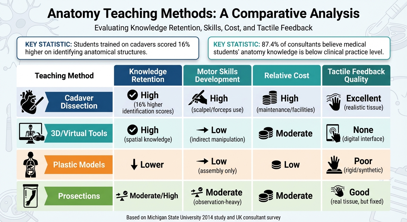
Research backs this up. A 2014 study from Michigan State University compared cadaver-based learning with the Anatomy and Physiology Revealed (APR) multimedia system. The results? Students trained on cadavers scored 16% higher on identifying anatomical structures and 11% higher on explaining how body parts function when tested on real cadavers.
"When it comes to learning actual – rather than simulated – human anatomy, the digital representations, despite their enhancements, did not work as well as the cadaver." – Cary Roseth, Associate Professor of Educational Psychology, Michigan State University
One of the key advantages of cadavers is the tactile feedback they provide. Students can feel the texture, resistance, and structure of real tissue - something no virtual tool or plastic model can imitate. While digital tools excel at showing processes like blood flow, they lack the physical interaction necessary for developing motor skills. Similarly, plastic models are rigid and fixed, limiting the realistic manipulation that cadaver dissection allows. This hands-on experience helps students better encode and retain complex anatomical knowledge.
Learning Outcomes Comparison
| Teaching Method | Knowledge Retention | Motor Skills Development | Relative Cost | Tactile Feedback Quality |
|---|---|---|---|---|
| Cadaver Dissection | High (16% higher ID scores) | High (use of scalpel/forceps) | High (maintenance/facilities) | Excellent (realistic tissue) |
| 3D/Virtual Tools | High (spatial knowledge) | Low (indirect manipulation) | Moderate | None (digital interface) |
| Plastic Models | Lower | Low (assembly only) | Low | Poor (rigid/synthetic) |
| Prosections | Moderate/High | Moderate (observation-heavy) | Moderate | Good (real tissue, but fixed) |
Another major benefit of cadaver-based learning is exposure to real anatomical diversity. Each cadaver is unique, showcasing variations in size, shape, and even pathology. This diversity mirrors what students will encounter in clinical practice, providing a level of preparation that standardized models and idealized digital tools cannot match. In fact, a study in the UK revealed that 87.4% of consultants believed medical students' anatomy knowledge was below the level needed for clinical practice.
Cadaver training stands out as the most effective method for preparing students to handle the complexities of real-world medicine. From tactile learning to exposure to natural variations, it offers an irreplaceable foundation for future clinicians.
Using Cadavers in Today's Anatomy Education
Combining Multiple Teaching Methods
Modern anatomy education takes a hybrid approach, blending traditional cadaver-based learning with advanced digital tools, 3D software, and radiological imaging. This combination leverages the strengths of each method to create a more comprehensive learning experience, a topic we explore in more depth in Bridging Tradition and Innovation in Anatomy Education.
Take the University of Heidelberg's Institute of Anatomy and Cell Biology, for example. They provided 50 students with access to both physical cadavers and life-sized CT images. The result? Students who studied using both resources saw their average test scores jump from 17.1 to 21.8 - a 27% improvement.
"Simultaneous physical and virtual dissection provide unique conditions to study anatomy." – Daniel Paech, Department of Radiology, German Cancer Research Center
Similarly, at University College Cork’s Department of Anatomy and Neuroscience, a rotation system introduced students to various learning stations. These included faculty-led cadaver prosections, plastic models, plastinated specimens, and digital tools like Acland's Video Atlas. Surveys conducted between 2018 and 2021 revealed that students overwhelmingly preferred cadaveric prosections paired with clinical tutorials, giving them a mean ranking of 1.40 out of 6. In contrast, plastic models ranked the lowest, with a mean score of 4.51 out of 6.
While digital tools excel at identifying structures and reviewing spatial relationships, cadavers offer tactile feedback and expose students to natural anatomical variations. This combination not only improves short-term learning but also lays the groundwork for clinical proficiency in the future.
Long-Term Benefits for Students
The value of cadaver-based learning goes far beyond test scores, deeply influencing clinical skills and professional confidence.
At the University of Colorado School of Medicine, Cadaver Review Sessions focused on peripheral nerve block anatomy had a significant impact. In February 2020, 98% of residents recommended incorporating these sessions into clinical rotations, citing improved anatomical understanding and enhanced clinical skills.
Cadaver training is also highly regarded by students in medical and dental programs. In one survey, 95% of medical students and 98% of dental students reported that cadaver dissection was beneficial to their learning. Meanwhile, 94% of medical students and 86% of dental students opposed replacing dissection entirely with computer-based alternatives.
"Human material remains the most superior way to teach anatomy and help achieve the biggest number of learning outcomes." – Joy Y. Balta, Department of Anatomy and Neuroscience, University College Cork
The long-term benefits of cadaver-based learning become especially clear when students enter clinical practice. Each cadaver presents unique anatomical variations - like differences in vessel branching, nerve pathways, and tissue structures - that prepare students for the unpredictability of real-world patient care. Unlike standardized digital models, cadavers offer an irreplaceable depth of understanding that directly translates to better clinical readiness.
Conclusion
Studies show that students who train with real human tissue gain a deeper understanding of spatial relationships, retain knowledge more effectively, and feel more confident in clinical settings compared to those using only plastic models or digital tools. For instance, 94% of surgical residents believe cadaver-based programs improve their anatomical education, and 53% report feeling more prepared to perform surgeries after this type of training. These benefits lay the groundwork for developing strong clinical skills.
Working with cadavers provides tactile feedback, highlights natural anatomical differences, and creates an emotional connection to learning that digital simulations simply cannot replicate. This hands-on experience exposes students to the variability of human anatomy, such as differences in vessel branching, nerve pathways, and tissue structures, preparing them for the complexities they’ll encounter in real-world clinical practice.
As previously discussed, these practical skills directly enhance surgical and diagnostic performance. Medical students who train with cadaveric material build the spatial awareness and technical abilities essential for their careers, contributing to better outcomes for their future patients.
FAQs
Why are cadavers better than digital tools for learning anatomy?
Cadavers offer an irreplaceable, hands-on learning experience that digital tools just can't match. They give students the chance to explore the three-dimensional structure of the human body, feel the texture of different tissues, and see natural anatomical variations up close. This kind of interaction is essential for understanding spatial relationships and getting ready for real-world clinical practice.
Research shows that working with cadavers leads to better knowledge retention and improved test performance compared to relying solely on digital simulations. While technology can be a great supplement, the tactile experience and authentic environment provided by cadavers remain unparalleled.
At the Institute of Human Anatomy, students get the best of both worlds. Real human cadavers are a key part of the curriculum, allowing learners to engage in hands-on dissection while also using modern multimedia tools. This blended approach helps students build a solid understanding of anatomy while taking advantage of cutting-edge educational resources.
How does learning with cadavers improve anatomy skills and confidence?
Cadaver-based learning offers a unique, hands-on opportunity to understand human anatomy in a way that models or screens simply can't match. Working with real human specimens allows students to feel the texture, elasticity, and spatial relationships of tissues firsthand. This direct interaction is invaluable for practicing critical skills like suturing, vessel ligation, and nerve identification in a setting that closely mirrors real-life scenarios. It also sharpens accuracy, procedural techniques, and the ability to recognize anatomical variations - essential for effective patient care.
But it’s not just about technical skills. These sessions also build confidence. Students often report feeling more prepared for clinical tasks and exams after working with cadavers. By linking anatomy to practical, real-world situations, this experience enhances decision-making and readiness for patient care. The Institute of Human Anatomy incorporates these practices into its offerings, such as interactive labs and guided dissections, ensuring students are thoroughly equipped for clinical success.
What emotional benefits do students gain from learning with cadavers?
Learning with real human cadavers offers students more than just a lesson in anatomy - it provides an opportunity for personal and emotional growth. This hands-on experience pushes students to confront mortality, fostering a deeper sense of empathy, respect, and appreciation for the human body. Many students find that this process eases their fears about death and strengthens their commitment to compassionate patient care.
Beyond that, working with cadavers helps students build emotional resilience and confidence. Facing the intimate and often challenging realities of dissection prepares them for the emotional complexities of clinical practice. It shapes their professional identity with a foundation of compassion and humility, qualities essential for meaningful patient care.

