
Wearable Tech vs. Cadaver Labs: Key Differences
Dec 19, 2025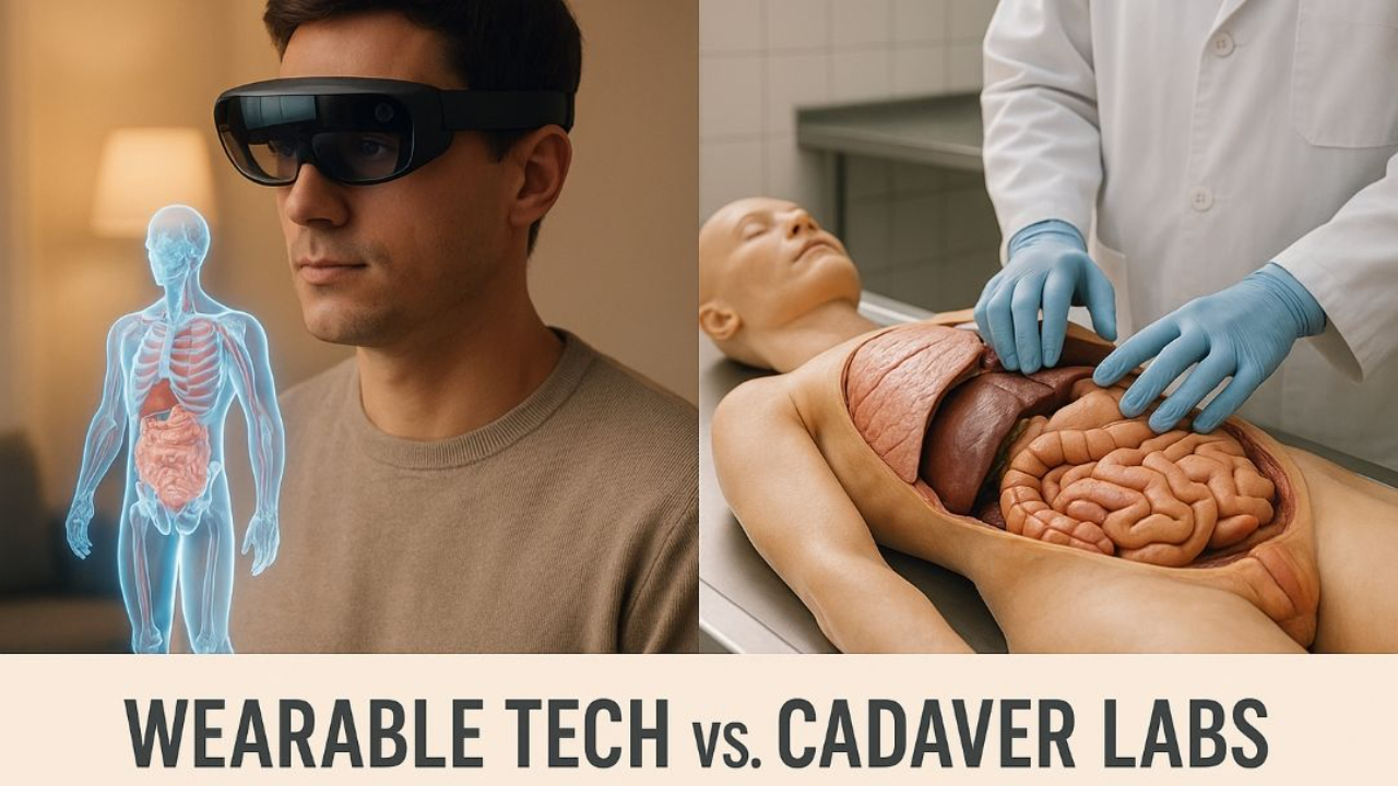
Medical education is shifting between two approaches: wearable technology and cadaver labs. Both offer unique benefits for learning anatomy, but they serve different purposes:
- Wearable Tech: Tools like VR and AR provide 3D models, remote access, and unlimited practice opportunities. These are cost-efficient long-term and great for conceptual learning.
- Cadaver Labs: Offer hands-on experience with real human tissue, showing anatomical variations and building practical skills essential for clinical practice.
Key Takeaway: Wearable tech is excellent for accessibility and visualization, while cadaver labs provide irreplaceable tactile learning. Combining both methods creates a balanced educational approach.
Quick Comparison
| Factor | Wearable Technology | Cadaver Labs |
|---|---|---|
| Learning Style | Interactive 3D models, remote access | Hands-on dissection, real tissue |
| Cost | High initial cost, low ongoing expenses | High ongoing costs for facilities |
| Practice | Unlimited, self-paced | Limited by lab schedules |
| Anatomical Detail | Standardized models | Real variations in anatomy |
| Skill Building | Useful for basic understanding | Builds critical surgical skills |
For the best results, start with wearable tech for theory and move to cadaver labs for hands-on practice.
How VR is Revolutionizing Anatomy Education
Wearable Technology in Anatomy Education
Wearable technology is transforming anatomy education, offering students new ways to explore the complexities of the human body. Devices like VR headsets, AR glasses, and smartwatches provide opportunities to study anatomical structures in ways that were once unimaginable.
These tools create immersive environments where students can interact with detailed, manipulable 3D models of the human body. AR glasses, for example, can project text or graphics directly onto physical objects, enhancing real-world visualization. VR goes a step further, allowing learners to dissect virtual bodies, practice surgical techniques, and examine structures from every angle in a fully immersive space.
The functionality of wearable technology isn’t limited to head-mounted devices. Smartwatches and fitness trackers are also being used to monitor biometric data during training sessions. This real-time feedback helps students connect theoretical knowledge with practical concepts like vital signs and patient care, making the learning experience more dynamic and engaging.
Benefits of Wearable Technology
Research highlights several advantages of using wearable technology in anatomy education. For instance, 50% of studies on VR adoption have shown statistically significant improvements in knowledge retention and acquisition.
Here are some of the most notable benefits:
- Accessibility: Virtual dissections and digital lessons make high-quality anatomy education available to students in remote or underserved areas, removing geographic barriers.
- 3D Visualization: Students can manipulate anatomical models - rotating them, peeling back layers, and observing real-time functions. This hands-on approach deepens understanding of complex relationships within the body.
- Cost Efficiency: While the initial investment in devices like VR headsets and AR glasses may be high, they eliminate the recurring costs of cadaver labs. Virtual dissections can be repeated endlessly without additional expenses.
- Self-Paced Learning: Wearable technology allows students to revisit challenging topics as often as they need, without being tied to lab schedules or cadaver availability.
- Personalized Learning: Smart devices track engagement and comprehension, enabling educators to tailor lessons to individual needs. For example, studies show that wearable tech helped students with autism improve peer interactions by 30%, while reducing anxiety triggers for ADHD students by 20%.
- Connectivity: Wearable devices integrate seamlessly with other digital tools, creating a cohesive learning environment that complements traditional study materials and management systems.
Drawbacks of Wearable Technology
Despite its advantages, wearable technology does have limitations. One of the biggest challenges is the lack of tactile feedback. While virtual tools can simulate anatomical structures, they cannot replicate the physical sensation of handling real tissues, such as texture or resistance.
The upfront cost of hardware like VR headsets and AR glasses can also be a significant hurdle, especially for smaller programs with limited budgets. Although these costs may decrease over time, the initial investment remains a barrier for many institutions.
Another challenge lies in the learning curve. Both students and instructors need training to use these devices effectively, and not everyone adapts at the same pace. Additionally, the effectiveness of wearable technology can vary based on factors like VR setup, interaction methods, and user demographics. The data generated by these devices also requires expert interpretation to ensure that learning objectives are being met safely and effectively.
Common Applications
Wearable technology is already making a meaningful impact in anatomy education through a variety of applications. Virtual dissections, for instance, allow students to explore detailed anatomical structures without relying on cadavers. This eliminates many logistical and ethical barriers while providing an equally in-depth experience.
VR headsets are being used to simulate surgeries and medical procedures, offering students a safe, repeatable environment to practice and learn from mistakes without putting real patients at risk. AR technology, on the other hand, is particularly useful for teaching internal anatomy, such as the ear, nose, and temporal bone, as well as specialized fields like surgical, neuro-, and cardiac anatomy.
Remote training programs are another area where wearable technology shines. By leveraging these tools, institutions can provide anatomy education to students who might otherwise lack access to cadaver labs or other resources.
Wearable simulators also add a layer of realism to clinical training, bridging the gap between theoretical knowledge and hands-on practice. Medical students and residents have reported high levels of satisfaction and engagement with these technologies, finding them both enjoyable and effective. By blending physical and digital elements, wearable technology complements traditional cadaver-based methods, enriching the learning experience and enhancing outcomes in healthcare education.
Cadaver-Based Anatomy Education
Even with the rise of digital tools, cadaver labs remain a cornerstone of anatomy education, offering a hands-on experience that no screen can replicate. The process of dissecting and examining real human tissue provides future medical professionals with a deep understanding of anatomy, shaping how they diagnose and treat patients throughout their careers.
Cadaver-based learning stands apart from digital methods by exposing students to the true complexity of the human body. Real tissue reveals natural anatomical variations that are critical for clinical practice, preparing students for the reality that every patient is unique. This hands-on approach not only enhances understanding but also highlights the strengths and challenges of using cadavers in medical training.
Benefits of Cadaver Labs
Dissecting cadavers offers a tactile, immersive learning experience that digital tools simply can’t match. Students encounter anatomical variations - like organs that differ in size or blood vessels that follow unexpected paths - that textbooks rarely address. This exposure prepares them for the unpredictable nature of real-world patient care.
Cadaver labs also help students develop practical skills. Handling real tissue provides realistic feedback that sharpens surgical techniques, improves hand-eye coordination, and builds muscle memory. In this risk-free environment, students can practice complex procedures, make mistakes, and learn from them without jeopardizing patient safety. The result? Greater confidence and competence when they transition to working with living patients.
Beyond technical skills, working with cadavers instills a deep respect for the human body. It reinforces professional values like ethical patient care, accountability, and the privilege of practicing medicine. This emotional and psychological connection to their work stays with students long after their anatomy lessons, influencing their approach to healthcare.
Drawbacks of Cadaver Labs
Despite their value, cadaver labs come with significant challenges. High costs for maintaining facilities and procuring cadavers make them inaccessible to many institutions, especially in smaller programs or developing regions. This financial barrier creates disparities in anatomy education, leaving some students with limited or no access to cadaver-based training.
Cultural and ethical factors also limit cadaver availability. In many parts of the world, religious beliefs, cultural attitudes toward body donation, and the lack of formal donation programs lead to severe shortages. Even in regions with established donation systems, aging populations and declining donation rates can create supply issues. These challenges mean some medical students miss out on critical hands-on training, potentially affecting their readiness for clinical practice.
How Cadaver Labs Are Used Today
Despite these challenges, cadaver labs remain a vital part of medical education, evolving to meet modern needs. They provide essential training for undergraduates learning anatomy and advanced practice for surgical residents. Many surgical specialties even require cadaver-based training for certification, as it allows direct evaluation of skills like technique and decision-making.
Organizations like the Institute of Human Anatomy are expanding access to cadaver-based education. By combining hands-on cadaver work with digital tools, they teach anatomy and physiology to a wide audience, including students, educators, and healthcare professionals. This hybrid approach respects the unique value of cadaver labs while making the learning experience more accessible.
Today, many institutions blend cadaver labs with digital technologies like virtual and augmented reality. This integrated model allows students to build foundational knowledge through digital tools before applying it in hands-on dissection. By combining the strengths of both methods, schools can maximize learning outcomes while conserving limited cadaver resources.
For surgical specialties, cadaver-based training remains indispensable. It offers a level of realism and direct engagement that virtual simulations cannot fully replicate, ensuring that future surgeons are well-prepared for the complexities of their work.
Wearable Tech vs. Cadaver Labs: Side-by-Side Comparison
Choosing between wearable technology and cadaver labs for anatomy education depends on your learning goals. Each approach has its strengths, and understanding how they compare can help educators and students make informed decisions.
Comparison Table
| Factor | Wearable Technology | Cadaver Labs |
|---|---|---|
| Knowledge Retention | Studies show 50% improvement in knowledge retention with VR adoption | Multisensory experiences enhance memory through visual, tactile, and olfactory inputs |
| Accessibility | Accessible remotely, allowing learning from anywhere with internet access | Requires physical attendance at specialized facilities, limiting reach |
| Cost Structure | Initial investment in hardware (e.g., VR headsets, AR glasses) and software licensing avoids ongoing costs | High ongoing costs for facility maintenance, cadaver procurement, and ethical oversight |
| Learning Modality | Excels in conceptual understanding and 3D visualization of complex structures | Offers tactile feedback and hands-on dissection experience with real human tissue |
| Scalability | Can serve many students simultaneously across multiple locations | Limited by space, cadaver availability, and facility capacity |
| Practice Frequency | Allows unlimited, self-paced practice without time constraints | Restricted by scheduled lab sessions and cadaver availability |
| Student Engagement | Immersive environments and gamification enhance satisfaction | Direct interaction with human donors fosters emotional connection and professional identity formation |
| Safety | Provides a risk-free environment for practicing procedures | Requires strict biohazard protocols and infection control measures |
| Anatomical Variation | Standardized anatomy with limited exposure to natural variations | Displays authentic anatomical differences critical for clinical practice |
| Skill Development | Ideal for initial skill-building and repeated practice | Essential for refining clinical judgment and adaptability with real tissue |
Wearable tech shines in accessibility and scalability, while cadaver labs deliver unparalleled hands-on experience.
Which Method Works Better for Different Learning Goals
The choice between wearable technology and cadaver labs often depends on the specific learning objective.
For building foundational knowledge, wearable technology is incredibly effective. Its 3D visualization tools allow learners to explore anatomical structures from various angles and revisit material as needed. This is especially helpful for students in remote areas, where virtual dissections remove geographic barriers and offer safe, repeatable practice.
When it comes to developing procedural skills and surgical expertise, cadaver labs are irreplaceable. Handling real tissue helps students build muscle memory and refine clinical judgment - skills that simulations alone can't fully replicate.
Wearable technology also promotes inclusivity in education. For example, studies show that VR and AR tools can improve learning experiences for students with special needs. In one case, students with autism saw a 30% increase in positive interactions, while teachers reduced anxiety triggers for students with ADHD by 20%.
The Institute of Human Anatomy highlights how blending cadaver-based education with digital tools can enhance learning. By integrating technology, traditional cadaver labs can reach a broader audience while maintaining their hands-on advantages.
Ultimately, the best approach may be a combination of both methods. Digital tools are excellent for introducing concepts and offering flexible access, while cadaver labs remain essential for developing fine motor skills and clinical expertise. A blended strategy - starting with virtual tools and transitioning to hands-on practice - can provide a well-rounded anatomy education.
Virtual reality, augmented reality, and other digital tools have proven to be effective supplements to cadaver-based learning, particularly when access to physical specimens is limited. These technologies aren't replacements but complementary tools that serve distinct educational purposes. Together, they create opportunities for innovative, mixed learning approaches that maximize the benefits of both methods.
Combining Wearable Technology and Cadaver Labs
The best anatomy education doesn’t pit wearable technology against cadaver labs - it combines them. By integrating both, students can achieve deeper, more effective learning outcomes.
Mixed Learning Approaches
Integrated approaches leverage the strengths of both wearables and cadaver labs, creating a richer educational experience.
Start with wearables, then move to cadavers: Using VR or AR wearables as an introduction helps students build foundational knowledge before they encounter cadavers. This step-by-step approach addresses a common issue: the initial shock or overwhelm many students feel when working with cadavers for the first time.
One popular method is the "flipped lab" model. Students use VR headsets to explore anatomy and learn key terms and spatial relationships before stepping into the lab. By the time they work with cadavers, they’re ready to focus on hands-on skills and clinical applications rather than basic identification.
AR glasses during dissections bring a new layer of interactivity. These glasses overlay digital information directly onto cadavers, making structures like nerves and blood vessels visible in ways they wouldn’t be otherwise. This bridges the gap between theoretical knowledge and physical practice.
Smartwatches and other biometric wearables also add value by monitoring student stress levels during cadaver labs. For instance, data shows that these tools reduce anxiety triggers by 20% for students with ADHD. This real-time feedback allows educators to provide targeted support, creating a more inclusive and supportive environment.
New Developments in Anatomy Education
Emerging tools are pushing the boundaries of how digital technology and traditional dissection can work together.
Haptic feedback controllers allow students to feel tissue resistance, organ textures, and anatomical structures during VR practice. This prepares them for the tactile experience of cadaver dissection, letting them practice cutting techniques and organ manipulation in a safe, virtual environment.
Advanced haptic suits take this concept even further. These full-body systems simulate spatial relationships and movements during virtual procedures, offering a more immersive and realistic experience. Though still in development, they hold promise for replicating the feel of cadaver work.
Mixed reality systems combine physical cadavers with digital overlays. Students can examine real specimens while viewing additional details about internal structures, abnormalities, or functional relationships not visible to the naked eye. This blend of physical and digital learning creates a more comprehensive understanding of anatomy.
Hand-tracking controllers in VR are also evolving. These tools now allow natural hand movements during virtual dissections, helping students practice realistic techniques and build muscle memory that translates to actual cadaver work.
Biometric wearables are becoming even more advanced. Beyond monitoring stress, they now assess cognitive load, allowing educators to adjust the intensity of lessons. This ensures students are pushed to learn without feeling overwhelmed, whether in virtual or physical settings.
The benefits of these technologies are backed by research. Studies show that 50% of VR-based anatomy education programs (4 out of 8) report measurable improvements in knowledge retention and understanding. Students also report greater confidence and a stronger grasp of complex concepts when these tools are part of their training.
Collaboration and Implementation
For these mixed approaches to succeed, collaboration between technology experts and anatomy educators is critical. By combining their expertise, they can create learning frameworks that are both effective and sustainable, preparing students for real-world clinical challenges.
Institutions looking to adopt these methods should start small. Pilot programs using affordable AR applications can demonstrate the value of these tools before scaling up. Focusing on areas like neuroanatomy or cardiac anatomy - where 3D visualization makes the biggest impact - can maximize early success. From there, programs can expand based on measurable outcomes and student feedback.
Conclusion
Wearable technology and cadaver labs work hand in hand to enhance anatomy education. Together, they create a well-rounded learning experience by blending the strengths of digital tools with the irreplaceable value of hands-on dissection. Knowing how and when to use each method allows students and educators to maximize their potential.
Main Points
Wearable technology provides accessibility and immersive 3D visualization. These tools are especially useful for students who lack access to cadaver labs due to location or limited resources. They also offer flexible, personalized learning opportunities that cater to individual needs. Virtual platforms enhance understanding and build confidence in grasping complex concepts.
Cadaver labs deliver hands-on experience critical for clinical practice. Real tissue provides tactile feedback and spatial awareness that simulations simply cannot replicate. Students also gain exposure to natural anatomical variations and develop professional attitudes through the ethical considerations involved in working with human remains.
Combining both methods leads to better results. Studies reveal that 50% of research on VR adoption shows measurable improvements in knowledge retention and acquisition. By using both approaches, students gain a deeper understanding - benefiting from the standardized models in virtual tools and the real-world variability found in cadaver specimens.
Each method serves specific learning goals. Wearable technology is ideal for visualizing concepts, repeated practice, and remote learning. Cadaver labs, on the other hand, are essential for honing surgical skills, understanding tissue properties, and experiencing anatomical diversity. Many effective programs begin with VR or AR to establish foundational knowledge, then transition to cadaver labs for hands-on skill development.
These insights can help shape a more effective anatomy education strategy.
Next Steps
Use these principles to tailor your anatomy training to your goals and resources. Whether you're an individual learner or part of an institution, blending both approaches can elevate your understanding of human anatomy.
For those focusing on cadaver-based education, the Institute of Human Anatomy offers interactive courses that integrate real cadaver training with digital tools. With a massive following of over 24 million subscribers and more than 2 billion video views, they highlight the value of hands-on learning. They also feature AI Jonathan, an AI-powered assistant available 24/7 to simplify complex anatomy topics, provide study support, and answer questions.
For those exploring wearable technology, start by identifying the areas of anatomy you find most challenging. Regions like neuroanatomy and cardiac anatomy often benefit the most from 3D visualization. If you're limited in accessing cadaver labs, VR and AR tools offer an effective alternative for building a strong foundation.
The future of anatomy education lies in leveraging the strengths of both methods. Whether you begin with wearable technology to build confidence or use virtual tools to reinforce what you've learned in the lab, combining these approaches ensures a comprehensive understanding of human anatomy.
FAQs
How does wearable technology support students with special needs in learning anatomy?
Wearable technology offers a game-changing approach to anatomy education, especially for students with special needs. Tools like augmented reality (AR) glasses and haptic feedback devices allow learners to explore and interact with anatomical structures in real-time. This hands-on, interactive experience makes it easier to understand complex concepts while keeping the learning process engaging.
What’s more, these devices can be adjusted to meet individual learning needs. For instance, tactile feedback can assist students with visual impairments, while visual overlays can help those with auditory processing difficulties by reinforcing important ideas. By providing an immersive and flexible learning experience, wearable technology addresses challenges often faced in traditional teaching methods, making education more inclusive and effective.
What ethical considerations and challenges come with using cadaver labs for medical training?
Cadaver labs are a cornerstone of medical education, offering invaluable hands-on learning opportunities. However, they come with significant ethical considerations and challenges. A primary concern is honoring the consent and wishes of those who donate their bodies. These contributions must be handled with the utmost respect and used strictly for the educational purposes they intended.
Another layer of complexity lies in the emotional toll on students. Working with human cadavers can be deeply impactful, often requiring thoughtful guidance to help students navigate the emotional weight of the experience while fostering a sense of respect for the individuals who made this learning possible. Additionally, proper care in handling, maintaining, and ultimately managing the disposition of cadaveric materials is crucial to ensure all actions align with both ethical principles and legal requirements.
How can wearable technology and cadaver labs work together to enhance anatomy education?
Wearable technology and cadaver labs each bring distinct advantages to anatomy education, and combining them offers a more complete learning experience. Wearable tech introduces interactive tools like augmented reality (AR) visualizations and real-time feedback, making it easier to grasp complex systems in an engaging, dynamic way. It allows students to learn on the go, providing flexibility and accessibility.
On the other hand, cadaver labs deliver an unmatched opportunity to study real human anatomy. They offer the chance to explore physical structures and variations that digital tools can't fully replicate, giving students a deeper understanding of the human body through hands-on experience.
When used together, these methods complement one another perfectly. Wearable technology enhances accessibility and precision, while cadaver labs provide the tactile, real-world learning that’s essential for a comprehensive understanding of anatomy. This blend equips students and professionals with both theoretical knowledge and practical skills.

