
How 3D Cadaver Imaging Transforms Anatomy Learning
Mar 29, 2026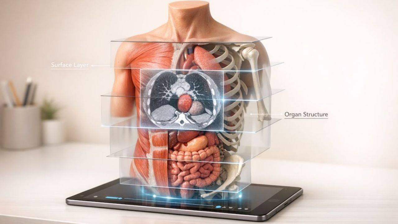
3D cadaver imaging is changing how anatomy is taught. By combining real human cadavers with interactive digital models, it offers a way to study anatomy without the limitations of traditional dissection labs. Here's why this matters:
- Accessibility: Students can explore detailed 3D models anytime, without needing physical specimens.
- Precision: Models are created using high-tech tools like CT scans and surface scanners, showing structures as small as 0.05 mm.
- Repeatability: Unlike dissections, digital models allow unlimited practice and reset options.
- Improved Learning: Many studies show that 3D tools can improve spatial understanding, short‑term test scores, and in some cases long‑term retention, though results vary by topic, learner group, and how the technology is integrated into teaching.
- Cost-Effective: Reduces expenses tied to maintaining cadaver labs, like preservation chemicals and ventilation systems.
These platforms also bridge the gap between anatomy and clinical imaging, helping students connect what they learn to real-world medical scenarios. With features like virtual dissection, physiological simulations, and integration with radiology data, 3D imaging is becoming a key tool for students, educators, and healthcare professionals alike.
Course: Cadaveric Images
How 3D Cadaver Imaging Works
Comparison of 3D Cadaver Imaging Technologies: CT/MRI, Serial Sectioning, Photogrammetry, and Surface Scanning
Creating digital cadavers combines cutting-edge imaging techniques to capture the surface and internal structures of human specimens. This process unfolds in three key stages: collecting data from real cadavers, digitally reconstructing that data into models, and transforming those models into interactive learning tools.
Data Collection from Real Cadavers
Building a 3D cadaver model starts with gathering detailed data from actual human specimens. Several technologies come into play here, each contributing unique insights.
- CT and MRI scans: These generate volumetric datasets in DICOM format, revealing internal structures and anatomical variations.
- Serial sectioning: In this method, cadavers are frozen and milled at 40-micron intervals, with each slice photographed in high resolution. Since 2004, Anatomage has documented over 2 million anatomical structures using this approach.
- Surface scanning: Handheld devices like the Artec 3D Spider capture surface details with sub-millimeter precision, achieving a 3D point accuracy of 0.05 mm and a mesh resolution of 0.1 mm.
- Photogrammetry: This technique involves taking hundreds of 2D photos from various angles - typically 196 images in about 15 minutes. Software then matches points across the images to form a 3D point cloud and surface mesh.
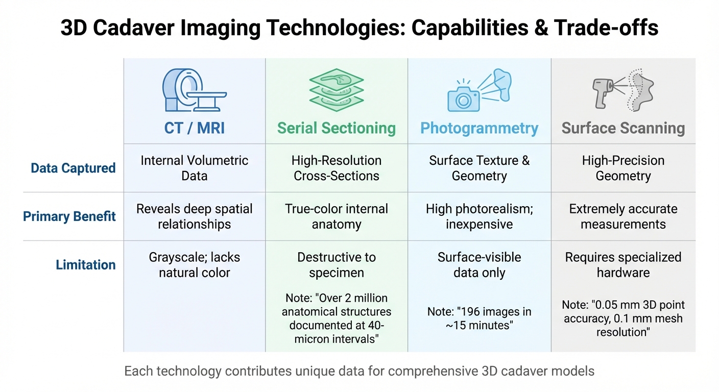
Each method has its strengths and limitations, as shown below:
| Technology | Data Captured | Primary Benefit | Limitation |
|---|---|---|---|
| CT / MRI | Internal Volumetric Data | Reveals deep spatial relationships | Grayscale; lacks natural color |
| Serial Sectioning | High-Res Cross-Sections | True-color internal anatomy | Destructive to specimen |
| Photogrammetry | Surface Texture & Geometry | High photorealism; inexpensive | Surface-visible data only |
| Surface Scanning | High-Precision Geometry | Extremely accurate measurements | Requires specialized hardware |
Once the data is collected, the next step is to refine it into detailed digital models.
Digital Reconstruction and Modeling
Turning raw data into a 3D model involves advanced software and skilled human input.
The process begins with image processing, which includes resampling, removing background noise, and adjusting colors to correct artifacts. Following this, segmentation is performed to identify and label specific structures like organs, blood vessels, and nerves. This step combines automated tools with manual tracing to account for unique anatomical features and pathologies.
Finally, volumetric rendering converts cross-sectional images into fully interactive 3D visualizations. Anatomage’s software achieves a resolution of 0.2 mm, enabling intricate details to be displayed.
"Anatomage's real cadaver content is the most unique on the market... exclusive slice cadaver data sets can go as low as 0.2 mm resolution, which allows visualization of much more detailed human anatomy."
– Anatomage
Interactive Learning Tools and Platforms
The final stage involves turning static models into dynamic tools for education. These interactive features make the models come alive for students and professionals.
Virtual dissection tables allow users to rotate and zoom in on models, toggle between transparent and full-tissue views, and explore the relationships between surface and internal anatomy. Unlike traditional dissection, these tools let students reset and repeat exercises as needed, without worrying about material limits.
Some platforms take this further by integrating physiology simulations. These features show movements like a beating heart, muscle contractions, and neural activity, helping learners connect anatomy to real-life function. This dynamic approach enhances spatial understanding and bridges the gap between academic knowledge and clinical practice.
In September 2025, researchers from Jagiellonian University Medical College in Kraków, Poland, showcased this process. Led by Weronika Michalik and a team of 13 experts, they used an Artec 3D Spider scanner with Artec Studio 17 Professional software to digitize eight cadaver specimens. Their work produced 12 detailed 3D models of organs, including the brain, heart, and pelvis, finalized using Blender 4.2.3 LTS software for interactive education.
"Anatomy is fundamentally a three-dimensional subject, and the benefits of three-dimensional learning tools are now undeniable and widely recognized."
– Weronika Michalik, Department of Anatomy, Jagiellonian University Medical College
Educational Benefits of 3D Cadaver Imaging
Improving Spatial Understanding and Retention
For many medical students, translating 2D textbook images into a 3D mental map is a tough hurdle, often leaving gaps in their anatomical knowledge. 3D cadaver imaging eliminates this challenge by letting students interact with digital models in three dimensions. This hands-on approach transforms abstract anatomical concepts into something tangible and easier to grasp.
In one study, students using the Anatomage Table improved their test scores by about 27% compared with a control group; across studies, score improvements vary depending on the curriculum and assessment. Similarly, recent meta‑analysis found that learners using 3D‑printed models achieved modestly higher test scores than those using traditional methods, with moderate effect sizes on average, though the precise percentage gain varies by study and topic. When studying intricate structures like the middle ear, students using 3D models achieved scores of 65.1%, outperforming those taught through conventional methods.
Stereo CT technology takes it a step further by combining traditional scans with stereoscopic visualization, significantly enhancing depth perception. Modern platforms can display sub‑millimeter details (around 0.5 mm) and provide clear, manipulable 3D views that are difficult to achieve with flat illustrations and sometimes more convenient than repeated cadaveric dissection, especially for deep or complex regions. This is particularly useful for studying hard-to-access structures like the middle ear, pterygopalatine fossa, or complex arterial systems, which often remain elusive in traditional methods.
By making these structures more accessible and understandable, 3D imaging paves the way for more engaged and independent learning.
Supporting Active and Self-Directed Learning
3D digital models revolutionize how students engage with anatomy. Unlike physical cadavers, which degrade with repeated use, 3D platforms offer endless opportunities for "dissection" without any material limitations. This repeatability is key for mastering complex relationships. In one survey, about 82% of students felt that working with 3D models improved how well they remembered anatomy over time. While many studies report perceived gains in understanding and satisfaction, objective evidence for long‑term retention is still limited and context‑dependent.
The benefits extend to academic performance. 86% of studies found that virtual dissection tables helped boost test scores, with gains ranging from 8% to 31% compared to traditional methods. Take the example of the All India Institute of Medical Sciences (AIIMS) in Bathinda: in December 2024, they introduced a 3D atlas to their dissection labs for 91 first-year MBBS students. The results? 97% of students reported better understanding and orientation of anatomical structures, and many noted increased enthusiasm and participation in the lab.
Interactive features like zooming, rotating, and peeling away layers keep students actively involved rather than passively observing. These tools are highly effective, with 73% of students finding 3D imaging platforms helpful for enhancing active learning during practical sessions. Cloud‑based systems that run on mobile devices support self‑paced study and review outside scheduled labs. Early research suggests they can improve learning self‑efficacy and perceived understanding, though their specific impact on stress reduction and long‑term memory is still being studied.
"VDTs represent a valuable complement to traditional anatomy education, enhancing learning outcomes and student engagement across a range of healthcare disciplines." – BMC Medical Education
Connecting Anatomy and Clinical Imaging
3D imaging doesn’t just improve spatial understanding - it also bridges the gap between anatomical study and clinical imaging. Historically, students have struggled to connect what they learn in anatomy with what they see in CT or MRI scans. 3D cadaver imaging solves this problem by integrating real-world clinical datasets, showing students how anatomical structures appear in medical imaging. Commercial Virtual Dissection Tables allow users to switch between X‑ray‑like transparency and soft‑tissue views, deepening understanding of radiographic anatomy. Vendor reports indicate global deployment across many countries, though exact counts are based on company data rather than independent studies.
Digital libraries that include real‑world cases and radiology reports help students practice linking anatomical knowledge with imaging findings, and are expected to support later clinical reasoning, although long‑term diagnostic outcomes are not yet well studied. Advanced platforms even simulate physiological behaviors, such as heart motion, blood flow, and muscle movement, helping students form accurate "mental models" for interpreting 2D scans within a 3D framework.
A standout example comes from Jagiellonian University Medical College in Poland. In September 2025, researchers there developed 12 high-fidelity 3D models of human cadaveric specimens with 0.1 mm resolution and 0.05 mm accuracy. By pairing these models with 2D images, they created an interactive digital library that enhanced spatial comprehension for both students and clinicians, effectively linking theoretical knowledge with clinical application.
Key Features and Use Cases of 3D Cadaver Imaging Platforms
Building on how 3D cadaver imaging works and its educational benefits, this section dives into the standout features and practical ways these platforms are used.
Core Features of 3D Imaging Platforms
Modern 3D cadaver imaging platforms go far beyond simple digital models. For example, volumetric dissection allows users to peel back layers - skin, muscle, bone - revealing internal systems without ever needing a scalpel. Many platforms include thousands of annotated structures, enabling learners to pinpoint specific anatomical parts from detailed lists.
A standout feature is physiological simulation. Advanced systems simulate dynamic processes like heartbeats, muscle contractions, neural activity, and respiratory motion. On some platforms that integrate simulation or skill‑lab features, learners can practice selected procedures (for example, ultrasound guidance or basic surgical approaches) in a risk‑free digital environment. Availability and realism of these procedure simulations vary between systems. Additionally, users can filter by systems (vascular, skeletal, nervous) or focus on specific regions like the thorax or head for targeted exploration. The real-time slicing capability offers moving cross-sections, providing detailed internal views.
The precision of these platforms is astounding, capturing details as fine as 0.2 mm to 0.5 mm. Some commercial systems report mapping over 2 million labeled anatomical structures, reconstructed from frozen cadaver slices as thin as about 40 μm (roughly the thickness of a human hair), although these figures come from vendor documentation rather than independent audits. To add even more depth, many platforms include pathology and histology libraries with thousands of patient cases and over 1,100 digital histology scans, making it easy to compare healthy and diseased tissues.
These advanced tools open up a wide range of applications for students, educators, and professionals alike.
Applications for Students, Educators, and Professionals
Different user groups benefit from 3D imaging platforms in unique ways.
Students often use these tools to prepare for physical dissections, reviewing 3D structures to grasp spatial relationships before entering the cadaver lab. Digital dissections allow for repeated practice without the limitations of physical specimens, encouraging self-paced learning with user-friendly interfaces and built-in learning assistants.
Educators can simplify complex topics with immersive 3D visuals, which can even be projected for large classrooms. With access to over 1,000 preset anatomical cases, instructors can tailor lessons, create quizzes, and design practical exams directly within the platform. For group learning, it's suggested that four to six students share a life-size digital dissection table for maximum engagement.
Healthcare professionals use these platforms to explore complex anatomy and plan procedures, which can support diagnostic reasoning, although direct evidence for improved diagnostic accuracy is still emerging With 3D DICOM rendering, clinicians can analyze patient scans in detail. Surgeons can simulate procedures in a virtual environment to minimize risks, while large touchscreens - often 84 inches - help doctors explain conditions to patients in 3D, making complex medical concepts easier to understand.
For example, the University of Connecticut Health in 2025 integrated 16 student workstations with 4K PCs and advanced digital dissection systems, enabling a fully virtual anatomy lab experience. Similarly, Truman State University uses 3D imaging tables in small group settings, allowing students to explore organ systems through guided activities that complement traditional lectures.
"The Virtual Anatomy Lab will be used by so many departments across Tech campus - Biology, Nursing, Kinesiology - just to name a few. It's cutting-edge technology that helps position Tech as a premier university in the life sciences." – Justin Hinckley, Louisiana Tech University
This flexibility and accessibility set these platforms apart from traditional cadaver labs.
Comparison with Cadaver Labs
3D imaging platforms address many of the challenges associated with traditional cadaver labs, offering a modern and efficient alternative. While cadaver labs provide the tactile experience of working with real tissue, they come with notable drawbacks. The table below highlights key differences:
| Factor | Traditional Cadaver Lab | 3D Cadaver Imaging Platform |
|---|---|---|
| Realism | High (actual tissue) but static; no motion | High (digitized human) with animated physiology, but lacks real tissue feel. |
| Accessibility | Limited by lab hours and donor availability | 24/7 access; supports remote learning |
| Cost | High recurring costs (donors, chemicals) | High initial cost; lower recurring costs than cadaver labs but still requires maintenance and licensing. |
| Safety | Exposure to formaldehyde and sharp tools | Chemical-free; no scalpels required |
| Repeatability | One-time use; mistakes can't be undone | Unlimited practice; structures reset instantly |
| Environment | Requires ventilation; strong odors | Odor-free; works in standard classrooms |
| Clinical Link | Difficult to connect with live imaging | Integrates directly with DICOM/radiology data |
Traditional cadaver labs often face logistical challenges like donor shortages, high costs, and the need for specialized facilities. In contrast, 3D platforms provide a standardized experience, ensuring every student sees the same high-quality structures. They also eliminate concerns like formaldehyde exposure, ventilation requirements, and specimen disposal.
The Future of Anatomy Education and 3D Cadaver Imaging
Next‑generation 3D cadaver imaging is reshaping anatomy education by offering more immersive, accessible, and often more effective learning experiences when used to complement traditional methods.
Trends in 3D Imaging Technology
Advancements in technology are revolutionizing how anatomy is visualized and taught, blending innovation with established methods.
One major innovation is haptic (force) feedback. In some advanced simulators, learners can feel resistance or texture when interacting with virtual tissues, adding a hands‑on element to digital dissections. Haptic systems are most established in surgical simulation and remain relatively specialized in anatomy teaching.
AI-driven modeling is also making waves. Machine learning automates the segmentation of images, drastically reducing the time and effort needed to identify organs and structures in CT and MRI scans.
Meanwhile, virtual and augmented reality (VR/AR) tools are transforming classroom experiences. Programs like Case Western Reserve University's HoloAnatomy, which uses Microsoft HoloLens, let students interact with 3D body models in mixed-reality settings. Initially adopted during the pandemic, this program has continued to thrive. 3D printing produces physical models based on imaging or cadaver scans, offering a sustainable option to complement, and in some constrained settings partially substitute for, traditional dissection, though cadaveric teaching remains the gold standard in most curricula.
Still, there's a growing recognition of the importance of hybrid learning. For example, the Kirk Kerkorian School of Medicine at UNLV relied solely on virtual anatomy tools for eight years. However, in late 2023, they reintroduced physical cadavers because students needed the sensory input and complexity that only real human anatomy provides. As Omid Rahimi, Director of the Human Anatomy Program at UT Health Science Center at San Antonio, explains:
"Current virtual models still cannot fully replicate the tangible complexity of human anatomy".
These innovations collectively enhance digital learning while supporting institutions in creating well-rounded educational experiences.
The Role of the Institute of Human Anatomy

Amid these technological advancements, the Institute of Human Anatomy is bridging the gap between traditional cadaver-based education and modern digital tools. Co-founded by Jeremy Jones and Jonathan Bennion, the Institute uses real human cadavers to deliver hands-on anatomy and physiology training. Their approach blends in-person experiences with digital resources like interactive courses, videos, and study guides. By integrating these tools, they make anatomy education accessible to students, educators, and healthcare professionals, offering a well-rounded learning platform that combines the best of both worlds.
Conclusion
3D cadaver imaging is reshaping anatomy education by addressing barriers that have limited access and learning quality. This shift is especially important as anatomy instruction hours in U.S. medical schools have significantly decreased. Unlike traditional dissections, which are limited to single-use, 3D models offer students the chance to revisit and study complex structures repeatedly, allowing them to learn at their own pace. With high‑fidelity scanners providing sub‑millimeter anatomical detail, these digital tools can approach cadaveric precision for visual learning while eliminating exposure to formaldehyde, though they do not replace the full tactile and procedural experience of cadaver labs.
These tools don’t just make anatomy more accessible - they enhance how students learn. As Weronika Michalik and her team at Jagiellonian University Medical College explained:
"Anatomy is fundamentally a three-dimensional subject, and the benefits of three-dimensional learning tools are now undeniable and widely recognized".
By improving spatial understanding, these platforms bridge the gap between theoretical knowledge and clinical imaging. They also allow students to practice selected procedures in a risk‑free environment, which can improve specific technical skills and may help prepare them for later real‑world training.
This shift is redefining anatomy education. Platforms like the Institute of Human Anatomy demonstrate how combining digital tools with cadaver-based learning can create a more comprehensive approach. Co-founded by Jeremy Jones and Jonathan Bennion, the Institute offers interactive courses, videos, and study materials, making anatomy education accessible to students, educators, and healthcare professionals around the world. This hybrid model provides the benefits of hands-on learning alongside the scalability of digital resources.
Looking ahead, advancements like haptic feedback, AI-driven personalization, and AR/VR integration promise to take anatomy education even further. By blending physical and digital experiences, these platforms equip students to meet the demands of modern healthcare, embodying the evolution of anatomy learning.
FAQs
Is 3D cadaver imaging as accurate as real dissection?
3D cadaver imaging delivers incredibly detailed and precise visuals of human anatomy, allowing users to interactively explore structures layer by layer. This technology stands out for its clarity and ease of access, making it an excellent tool for studying complex anatomical details. However, it doesn’t entirely replace the hands-on experience of traditional dissection, where students gain a deeper understanding of tissue textures and spatial relationships through touch. Each approach offers distinct advantages, and together, they create a well-rounded foundation for anatomy education.
Do 3D cadaver tools replace cadaver labs or complement them?
3D cadaver tools add a new dimension to anatomy education by improving visualization and offering interactive learning experiences. That said, they can't entirely substitute the hands-on, multisensory experience of working with real cadavers. Dissection helps students build essential manual skills and a deeper understanding of spatial relationships within the body. Together, these methods create a well-rounded approach to learning anatomy.
What do I need (hardware/software) to use 3D cadaver imaging at home?
To explore 3D cadaver imaging from home, you'll need a reliable computer or tablet that can handle advanced 3D visualization software. A high-performance computer with a sharp display or a touchscreen device works best for engaging, interactive views. For added convenience, mobile apps like Human Anatomy Atlas 2023 offer detailed 3D models accessible on smartphones or tablets. Just make sure your device meets the software's system requirements to ensure smooth performance.

