
What Are Hernias? Understanding Ventral and Groin Anatomy
Nov 09, 2026By Jonathan Bennion, MPA-C, Co-Founder & Lead Anatomist, Institute of Human Anatomy
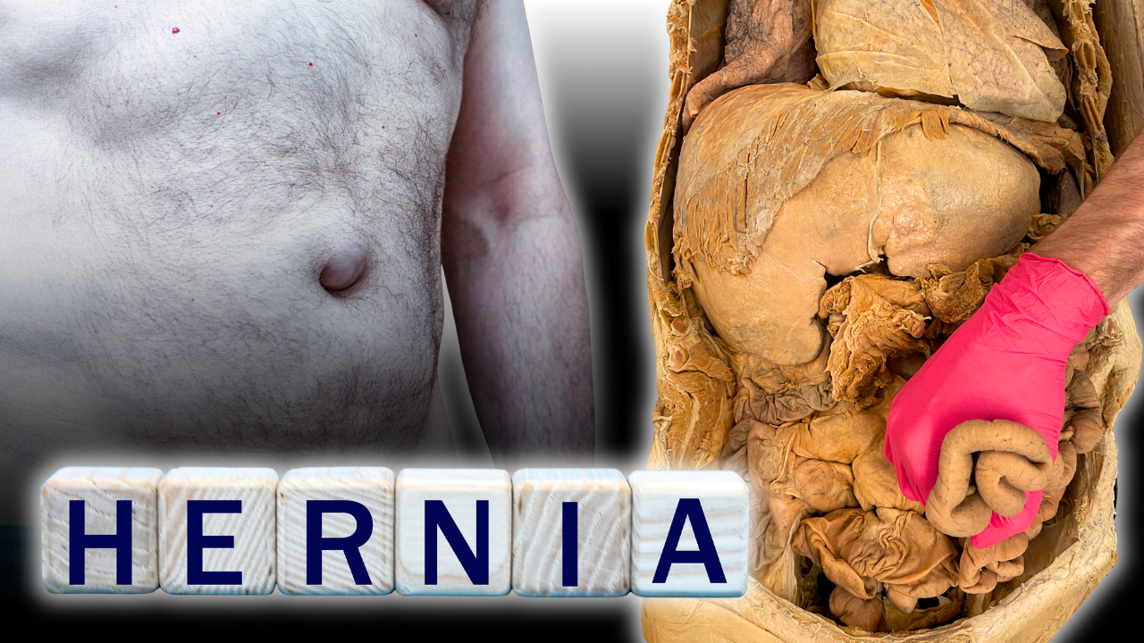
Hernias are common conditions that can affect different areas of the abdominal wall. They occur when an organ or part of an organ pushes through a weakened area of muscle or connective tissue, sometimes creating a visible bulge.
Understanding why hernias develop starts with looking at the anatomy of the abdominal wall and the structures that support it. Some hernias form along the midline of the abdomen, while others develop in the groin. These differences in location help explain why certain types are more common in men or women.
In this article, we'll look at the anatomy behind ventral and groin hernias, what can protrude through a weakened area, and what happens when a hernia leads to complications.
What Is a Hernia?
A hernia occurs when an organ or part of an organ protrudes through a weakened area in the abdominal wall. This can cause a noticeable bulge, particularly when standing, coughing, or straining.
Hernias are generally grouped into two main categories: ventral hernias and groin hernias. The main difference is where they develop.
Ventral hernias form along the front of the abdominal wall, often around the midline. Groin hernias develop in the lower abdominal region and include inguinal hernias and femoral hernias.
Ventral hernias are more common in women, while groin hernias, particularly inguinal hernias, are more common in men. The anatomy of the abdominal wall and differences in reproductive development help explain some of these patterns.
Ventral Hernias: Weakness Along the Midline
To understand ventral hernias, let's first look at the muscles and connective tissue along the front of the abdomen.
One of the most recognizable muscles in this area is the rectus abdominis, commonly called the six-pack muscle. It runs vertically along the front of the abdomen and is divided into sections by bands of connective tissue called tendinous inscriptions.
The rectus abdominis is surrounded by a layer of connective tissue known as the rectus sheath. Between the left and right rectus abdominis muscles is another important structure: the linea alba.
The term linea alba means "white line." It's a band of fibrous connective tissue formed by the joining of the aponeuroses, or flat tendons, of the abdominal muscles. It extends from the sternum down to the pubic bone.
Because the linea alba runs along the midline of the abdomen, weakness in this area can create an opening where a hernia may develop.
What Causes the Linea Alba to Weaken?
The linea alba can stretch, thin, or become weakened for several reasons. Some common contributing factors include:
- Pregnancy: As the abdomen expands during pregnancy, the abdominal wall stretches to accommodate the growing fetus. This can cause the linea alba to widen and the rectus abdominis muscles to move farther apart. This separation is known as rectus abdominis diastasis. Although diastasis recti is not a hernia itself, it can affect the strength of the abdominal wall and may occur alongside a hernia.
- Central Obesity: An accumulation of abdominal fat can place additional pressure on the abdominal wall. Over time, this pressure can contribute to stretching of the linea alba and separation of the rectus abdominis muscles.
- Congenital Factors: Some people are born with areas of weakness in the linea alba, which can make them more susceptible to developing a hernia.
Types of Ventral Hernias
When a weak spot develops along the linea alba, abdominal contents can push through it. The location of the opening determines the type of ventral hernia.
Umbilical Hernia
An umbilical hernia develops at or around the belly button, also called the umbilicus. This area is naturally weaker because of its role in fetal development, when the umbilical cord passes through the abdominal wall.
Umbilical hernias are common in infants, but they can also develop in adults, particularly when the abdominal wall is weakened or under increased pressure.
Epigastric Hernia
An epigastric hernia develops along the linea alba above the umbilicus. The epigastric region is the area between the sternum and the belly button.
Unlike an umbilical hernia, which forms at the navel, an epigastric hernia occurs elsewhere along the midline of the upper abdomen.
How Are Ventral Hernias Repaired?
Surgery may be recommended to repair a ventral hernia and reinforce the weakened area. Depending on the hernia, the surgeon may bring the connective tissue back together with sutures and use surgical mesh for additional support.
The mesh acts as reinforcement for the abdominal wall, helping reduce the chance of the hernia returning.
Groin Hernias: How Testicular Descent Affects the Anatomy
Groin hernias develop in the lower abdominal wall, near the junction between the abdomen and the thigh. The two main types are inguinal hernias and femoral hernias.
Inguinal hernias are much more common in men. One reason is related to how the reproductive organs develop before birth, particularly the descent of the testes.
During fetal development, both the testes and ovaries begin near the kidneys. As development continues, the ovaries descend into the lower pelvis, near the hip bones (os coxae).
The testes travel farther. They descend through the abdominal wall and into the scrotum. This process involves a passageway in the groin called the inguinal canal.
The pathway used by the testes creates an area of potential weakness in the male abdominal wall. This helps explain why men are more susceptible to inguinal hernias.
The Inguinal Ligament and Inguinal Canal
Two important structures help us understand groin hernias: the inguinal ligament and the inguinal canal.
The inguinal ligament runs from the anterior superior iliac spine (ASIS), a bony landmark on the hip, to the pubic bone. It's the structure that forms the boundary between the lower abdomen and the upper thigh. You may recognize it as part of the V-shaped crease in the groin.
The inguinal canal sits just above the inguinal ligament. During fetal development, the testes pass through this canal on their way into the scrotum.
As the testes descend, they bring several structures with them. These structures form the spermatic cord, which contains the testicular blood vessels (arteries and veins) and the vas deferens, the tube that carries sperm.
The inguinal canal provides a natural passage through the abdominal wall. However, this also makes it a potential site for weakness and hernia formation.
Types of Groin Hernias
Inguinal Hernia
An inguinal hernia develops when abdominal contents push through a weakened area in the inguinal region, often involving the inguinal canal.
Because of the anatomical pathway created during testicular descent, men have a greater risk of developing this type of hernia. A common sign is a bulge in the groin, which may contain part of the small intestine or other abdominal contents.
Femoral Hernia
A femoral hernia develops lower in the groin than an inguinal hernia. It occurs when abdominal contents pass through the femoral canal, just below the inguinal ligament.
Femoral hernias are less common than inguinal hernias, but they occur more frequently in women. They are still relatively uncommon compared with other types of hernias.
What Can Push Through a Hernia?
When an opening develops in the abdominal wall, different tissues and organs can push through it. Looking at a cadaveric specimen helps show what these hernia contents can include.
When the rectus abdominis is reflected, several structures can be seen near the abdominal wall that may contribute to the bulge of a hernia.
Preperitoneal Fat
Preperitoneal fat is fatty tissue located between the abdominal wall and the peritoneum, the membrane lining the abdominal cavity. In smaller hernias, this may be the only tissue that pushes through the weakened area.
Greater Omentum
The greater omentum is a large fold of fatty tissue that hangs from the stomach and drapes over the intestines. Part of it can push through an opening in the abdominal wall, particularly in larger ventral or umbilical hernias.
Small Bowel (Small Intestine)
Loops of the small intestine can also protrude through a hernia opening. This is a common finding, particularly in inguinal hernias.
Other possible contents include portions of the large intestine and the small fatty projections attached to its outer surface, known as epiploic appendages.
The contents of a hernia can vary depending on its location and size. What they have in common is that tissue or an organ has moved through an opening in the abdominal wall where it normally shouldn't be.
Hernia Complications: From Reducible to Strangulated
Not all hernias cause pain or require immediate surgery. Some remain small and cause few, if any, symptoms. However, certain complications can make a hernia a medical emergency.
Asymptomatic and Reducible Hernias
An asymptomatic hernia doesn't cause noticeable symptoms. A person may only become aware of it after noticing a small bulge in the abdomen or groin.
Some hernias are also reducible, meaning the protruding tissue can move back into the abdominal cavity, either on its own or with gentle pressure.
Whether a hernia needs surgical repair depends on several factors, including its size, symptoms, potential for complications, and the person's overall health. When surgery is performed, sutures and surgical mesh may be used to reinforce the weakened area.
Incarcerated and Strangulated Hernias
A more serious complication occurs when the tissue protruding through the hernia opening becomes trapped.
An incarcerated hernia is one in which the protruding tissue can no longer be pushed back into the abdominal cavity. The trapped tissue may cause pain and swelling, and the hernia may become increasingly uncomfortable.
If the blood supply to the trapped tissue becomes restricted, the hernia can progress to a strangulated hernia.
Strangulation occurs when the hernia opening puts enough pressure on the blood vessels supplying the trapped tissue to reduce or cut off its blood supply. Without adequate blood flow, the tissue can become damaged and eventually die. This is called necrosis.
A strangulated hernia is a life-threatening medical emergency. It requires immediate surgical treatment because the affected tissue can develop gangrene and lead to serious infection and other complications.
A new or worsening hernia bulge, especially when accompanied by significant pain, swelling, or other concerning symptoms, needs prompt medical evaluation.
The One Simple Habit That Will Prevent 90% of Groin Hernias
Key Takeaways: Understanding Hernia Anatomy
Hernias develop when organs or other tissues push through weakened areas of the abdominal wall. Their location helps determine the type of hernia and the anatomical structures involved.
Ventral hernias, such as umbilical hernias and epigastric hernias, often involve weaknesses along the linea alba. In the groin, the anatomy of the inguinal canal and the descent of the testes help explain why inguinal hernias are more common in men. Femoral hernias, which develop below the inguinal ligament, are more common in women.
Understanding these anatomical differences also helps explain what can protrude through a hernia opening and why complications such as incarceration and strangulation can occur.
Studying these structures through cadaveric dissection gives us a clearer view of how the abdominal wall is organized, where its natural weak points are, and how those areas relate to common hernia conditions.
Source: "What Are Hernias?" - Institute of Human Anatomy

About the Author
Jonathan Bennion, MPA-C, is Co-Founder and Director of Education at the Institute of Human Anatomy, where he has taught anatomy and physiology for over 15 years. A trained anatomist and lead dissector, Jonathan holds a Master of Physician Assistant degree and works as a medical provider in Urgent Care.

