
How Proprioception Guides Muscle Contraction
Apr 01, 2026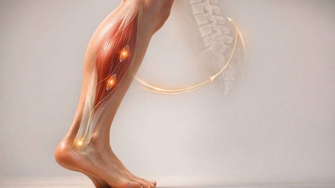
Proprioception, often called the "sixth sense", helps your body understand where your limbs are and how they move without needing to look. It’s powered by sensors in your muscles, tendons, and joints that send constant feedback to your brain. This system is crucial for smooth, controlled movements, balancing, and preventing injuries.
Here’s how it works:
- Muscle spindles detect muscle stretch and trigger contractions to maintain posture and prevent overstretching.
- Golgi tendon organs (GTOs) monitor muscle tension and can contribute to reflex pathways that modulate muscle activity, including inhibition, helping regulate force and protect tissues.
- Reflexes like the stretch reflex and mechanisms like alpha-gamma coactivation ensure precise and efficient muscle control.
- Proprioception adjusts muscle activity during both static (e.g., holding a plank) and dynamic (e.g., lifting weights) movements.
This feedback system operates automatically, allowing you to move without overthinking. Loss of proprioception, as seen in certain injuries or conditions, can lead to clumsy movements and higher injury risks. Training methods, such as balance exercises or proprioceptive neuromuscular facilitation (PNF), can improve this sense, making it vital for rehabilitation and athletic performance.
Proprioception is not just about movement - it underpins safety, coordination, and efficiency in most of your daily actions.
How Stretching REALLY Works
You can also learn human anatomy with real cadavers through our interactive courses and labs.
Main Proprioceptors in the Human Body
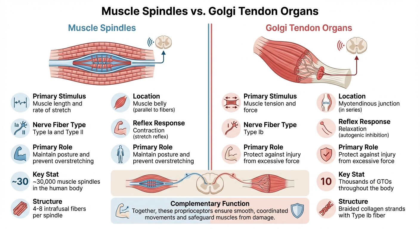
Muscle Spindles vs Golgi Tendon Organs: Key Differences in Proprioception
The body relies on tens of thousands of muscle spindles and many thousands of Golgi tendon organs to provide constant feedback for precise muscle control. Let’s break down how these two types of proprioceptors work and their unique contributions.
Muscle Spindles: Detecting Muscle Stretch
Muscle spindles are nestled within the belly of skeletal muscles, running parallel to the extrafusal fibers. Each spindle contains 4–8 intrafusal fibers wrapped in connective tissue. When a muscle stretches, these intrafusal fibers elongate, which deforms the sensory nerve endings. This deformation activates PIEZO2 ion channels, generating electrical signals that travel to the spinal cord.
"The major function of muscle spindles is to provide information about muscle length (that is, the degree to which they are being stretched)." - Purves D, et al., Neuroscience
Two types of nerve fibers handle this feedback: Type Ia fibers sense how quickly the muscle is stretching, while Type II fibers focus on the muscle’s static length. Interestingly, muscle spindles are largely absent in facial muscles and those of the middle ear.
The stretch reflex is a prime example of spindle function. Think of the classic knee-jerk test: when a doctor taps your knee, the sudden stretch activates muscle spindles, which then signal motor neurons in the spinal cord to contract the muscle. Gamma motor neurons play a supporting role by keeping the intrafusal fibers taut, ensuring the spindles remain responsive even during muscle contraction.
What Happens When You Stretch a Muscle!?
Golgi Tendon Organs: Monitoring Muscle Tension
While muscle spindles focus on length, Golgi tendon organs (GTOs) are all about sensing tension. These receptors are located at the myotendinous junction, where muscle fibers merge with tendons, and are arranged in series with the muscle fibers. They consist of braided collagen strands connected to a Type Ib sensory fiber.
When a muscle contracts, the force pulls on the tendon, compressing the collagen and activating PIEZO2 channels. This sends signals to the spinal cord about the tension being applied. GTOs are less sensitive to passive stretching since elastic fibers absorb most of the length changes. However, they respond strongly during active contractions, when tension directly impacts the tendon.
"If the muscle spindle system is considered a feedback system that monitors and maintains muscle length, then the Golgi tendon system is a feedback system that monitors and maintains muscle force." - Purves D, et al., Neuroscience
GTO input can participate in a reflex known as autogenic inhibition, which reduces muscle activity under some conditions and may help regulate high levels of tension, though its effects are more complex than a simple on–off protective switch. For example, during static stretching, sustained tension over several seconds can lead to reflex and mechanical changes that permit greater lengthening, contributing over time to improved flexibility.
Comparing Muscle Spindles and Golgi Tendon Organs
Here’s a quick side-by-side look at how these two proprioceptors differ:
| Feature | Muscle Spindles | Golgi Tendon Organs |
|---|---|---|
| Primary Stimulus | Muscle length and rate of stretch | Muscle tension and force |
| Location | Muscle belly (parallel to fibers) | Myotendinous junction (in series) |
| Nerve Fiber Type | Type Ia and Type II | Type Ib |
| Reflex Response | Contraction (stretch reflex) | Relaxation (autogenic inhibition) |
| Primary Role | Maintain posture and prevent overstretching | Protect against injury from excessive force |
Together, muscle spindles and Golgi tendon organs support smooth, coordinated movements and help protect muscles from excessive strain. Their complementary functions highlight the complexity of the body’s proprioceptive system.
Neural Pathways in Proprioception and Muscle Contraction
When proprioceptors detect changes in muscle length or tension, the nervous system springs into action, using specialized pathways to process this information and trigger the appropriate muscle response. These circuits work at lightning speed, bypassing conscious thought to produce reflexive movements. This intricate system explains how the body maintains balance, adjusts posture, and performs precise actions without us even realizing it. Let’s dive into the specific pathways that turn proprioceptive signals into coordinated muscle movements.
Stretch Reflex and Monosynaptic Pathways
The stretch reflex is one of the fastest neural pathways in the body. When a muscle spindle senses a sudden stretch, Group Ia sensory afferents send signals directly to the spinal cord. These signals form a direct connection with alpha motor neurons in the ventral horn, creating what’s known as a monosynaptic reflex arc.
"The excitatory pathway from a spindle to the α motor neurons innervating the same muscle is unusual in that it is a monosynaptic reflex" - Purves D, Augustine GJ, Fitzpatrick D, et al.
This single-synapse design is why the knee-jerk reflex (patellar reflex) happens so quickly. When the quadriceps are tapped, the muscle stretches, activating Ia afferents that connect directly with alpha motor neurons in the L2-L4 spinal segments. The result? An immediate contraction of the quadriceps and a forward kick of the leg. The stretch reflex is crucial for rapid postural adjustments and joint stability, with one of the shortest latencies of any spinal reflex.
Clinicians use this reflex to assess neurological health. Responses are graded on a scale from 0 (absent) to 4+ (hyperactive), with 2+ being considered normal. Abnormal responses can indicate damage to descending pathways or spinal circuits.
Alpha-Gamma Coactivation for Spindle Sensitivity
Reflexes alone aren’t enough for voluntary movement - spindle sensitivity must also be finely regulated. Here’s the challenge: when alpha motor neurons contract muscles, the spindles within those muscles would normally go slack, creating a sensory "blind spot." The body solves this with alpha-gamma coactivation, where the brain activates both alpha and gamma motor neurons at the same time.
While alpha motor neurons contract the main (extrafusal) muscle fibers to generate force, gamma motor neurons adjust the intrafusal fibers within the spindle. This keeps the spindle responsive, even during muscle contraction. Gamma motor neurons account for a substantial minority (roughly a quarter to a third) of the motoneuron fibers innervating a muscle, depending on the muscle and species. They have smaller axons (approximately 5 μm in diameter) and slower conduction speeds, ranging from 4 to 24 meters per second.
"Co-activation of the α and γ motor neurons allows spindles to function (i.e., send information centrally) at all muscle lengths during movements and postural adjustments." - Purves D, Augustine GJ, Fitzpatrick D, et al.
Gamma motor neurons come in two types: static gamma neurons, which adjust sensitivity to changes in muscle length (important for posture and slow movements), and dynamic gamma neurons, which enhance sensitivity to stretch velocity (critical for quick, reactive movements). The cerebellum and other supraspinal centers contribute to coordinating alpha and gamma activity, helping to regulate muscle tension during movement.
Reciprocal Inhibition for Coordinated Movement
Smooth motion isn’t just about contracting the right muscles - it’s also about relaxing the opposing ones. That’s where reciprocal inhibition comes into play. When Ia afferents from muscle spindles activate alpha motor neurons to contract a muscle, they simultaneously activate inhibitory interneurons in the spinal cord. These interneurons suppress the alpha motor neurons of the antagonist muscle, causing it to relax.
Take the knee-jerk reflex as an example. The same Ia afferents that contract the quadriceps also inhibit the hamstrings, ensuring they don’t resist the movement. This coordination allows for smooth, efficient motion.
Golgi tendon organs add another layer of control. When GTOs sense muscle tension, Group Ib afferents engage spinal interneurons that can inhibit the same muscle (often termed autogenic inhibition) and, in some cases, excite antagonists. These context‑dependent pathways help regulate force output and can contribute to joint stability.
Descending pathways, like the corticospinal tract, allow the brain to modulate these reflexes. This control lets us override reflexes when needed - like holding onto a hot dish - or amplify them in specific situations. It’s this interplay between reflexive and voluntary control that highlights the importance of proprioception in precise muscle coordination.
Proprioceptive Feedback in Muscle Control
Now that we've covered the neural pathways behind proprioceptive signals, let’s dive into how this feedback system manages different types of muscle contractions. Whether you're holding a yoga pose, lifting weights, or simply standing upright, proprioception fine-tunes muscle force in real time. This constant adjustment is what keeps us steady and coordinated, whether we're staying still or moving dynamically.
Isometric Contractions: Holding Steady
Isometric contractions occur when muscles generate force without changing length - like holding a plank or balancing on one leg. During these static activities, proprioceptive feedback is always at work, stabilizing your body. Muscle spindles detect subtle shifts in muscle length, while Golgi tendon organs (GTOs) monitor the force being applied. Together, they help maintain joint stiffness.
"Feedback from proprioceptive sensors also can directly contribute to muscle activity through reflexive loops to motor pools, potentially helping to maintain postural stability during body sway."
- Jesse C. Dean, Division of Physical Therapy, Medical University of South Carolina
This feedback system operates largely behind the scenes. For instance, if you’re standing and start to sway forward, muscle spindles in your calves sense the stretch and trigger a reflexive contraction to pull you back into balance. Meanwhile, the interplay between inhibitory signals from GTOs and excitatory signals from spindles ensures your joints remain stable through coordinated muscle contractions.
The importance of this system becomes glaring when it deteriorates. A June 2025 study by Łukasz Oleksy and Anna Mika at Wroclaw Medical University examined 48 active older women (average age 65 ± 6 years) performing isometric knee extensions at 75°. Participants aimed to reach 15%, 30%, and 60% of their maximum voluntary contraction without visual feedback. The study showed that actual muscle activation (measured by EMG) stayed below 10% for all targets, highlighting a disconnect between intended and actual muscle engagement that may reflect age‑related changes in proprioceptive and motor control. This decline helps explain why nearly one in three people over 65 experiences a fall each year, with hip fracture mortality rates reaching 20–30% within a year.
Isotonic Contractions: Moving with Control
Unlike isometric contractions, isotonic contractions involve movement - whether muscles are shortening (concentric) or lengthening (eccentric) under load. Here, proprioceptive feedback plays a key role in helping produce smooth, controlled motion. GTOs monitor the resistance your muscles face, adjusting force output accordingly.
Take the example of lowering a heavy box to the floor. This is an eccentric contraction, where your muscles lengthen under tension. During this process, muscle spindles track the rate of stretching, while GTOs monitor the increasing tension. If the load becomes very high, GTO input can increase inhibitory signaling to the spinal cord, reducing activation of that muscle and helping to regulate potentially excessive tension.
The precision of isotonic movements hinges on alpha-gamma coactivation. When muscles contract concentrically, gamma motor neurons activate intrafusal fibers in muscle spindles, keeping them taut and responsive. This ensures spindles continue providing feedback throughout the movement's full range. Without this coordination, our ability to perform precise, controlled actions would be severely impaired.
Force Modulation and Injury Prevention
Proprioceptive feedback doesn’t just guide movement - it also helps prevent injuries. When GTOs sense excessive muscle tension, they initiate an autogenic inhibition reflex, relaxing the muscle to avoid damage like tears or avulsions. Similarly, muscle spindles regulate stiffness, which determines how well muscles resist sudden joint movements. Higher muscle–tendon stiffness can reduce the mechanical delay between muscle activation and joint movement, which may support quicker mechanical responses to perturbations.
"Stiffer muscles should potentially resist sudden joint displacements more effectively... reducing the incidence of joint subluxation and injury."
- Bryan L. Riemann, PhD, ATC, University of Pittsburgh
Joint receptors provide another layer of protection. Acting like "limit detectors", they alert the nervous system when a joint nears its range of motion limits, helping prevent overextension or hyperflexion. This mechanism is crucial during high-impact activities like landing from a jump or making quick directional changes.
Proprioceptive input helps build internal models of body mechanics that support feedforward control, where the nervous system anticipates load changes and adjusts muscle activation in advance. For example, before stepping onto uneven ground, your body prepares your ankle muscles to stabilize against potential rolling. This anticipatory mechanism explains why proprioceptive training can enhance motor skills and reduce injury risk.
Practical Applications in Education and Training
Understanding proprioception isn’t just about theory - it’s about seeing how it works and applying it in real-world contexts. Education and training programs use these principles to bridge the gap between abstract concepts and practical skills.
Using Cadaver-Based Anatomy to Study Proprioception
Learning about proprioception in theory is one thing, but observing it in actual human tissue takes understanding to a whole new level. Cadaver-based anatomy education provides students and healthcare professionals with a hands-on opportunity to see the structures that make proprioception possible. For example, the Institute of Human Anatomy uses cadavers to show how muscle spindles are embedded within muscle bellies and how Golgi tendon organs (GTOs) are positioned at myotendinous junctions. This approach helps learners connect physiological concepts to tangible anatomical structures.
Dissecting the myotendinous junction reveals GTOs aligned with extrafusal fibers, offering a clear visual of their role. Students can also observe how muscle spindle density varies widely, on the order of a few up to around 100 or more spindles per gram in some muscles, across the body’s tens of thousands of total muscle spindles. Seeing these structures in person makes it easier to grasp how they detect stretch, tension, and joint position in real-time scenarios.
Improving Proprioceptive Awareness Through Training
This anatomical knowledge lays the groundwork for effective training techniques aimed at improving proprioceptive feedback. Whether someone is recovering from an injury or enhancing athletic performance, targeted exercises can sharpen proprioceptive skills. For example, balance training on unstable surfaces like wobble boards pushes the nervous system to rely more on internal signals rather than visual cues.
A February 2024 case report by Snehal S. Samal and colleagues highlights the impact of such training. A 45-year-old woman recovering from Achilles tendon reconstruction underwent 8 weeks of exercises, including wobble board and Swiss ball activities along with proprioceptive neuromuscular facilitation (PNF) techniques. Her Foot and Ankle Disability Index score improved from 20/104 to 98/104, and her pain level dropped from 6/10 to just 2/10 during activity.
"Proprioception allows for the programming of neuromuscular motor control, which is required for precise movement performance and the induction of muscle reflexes that maintain the joint's dynamic stability." - Samal et al., Neuro Physiotherapy, Datta Meghe Institute of Higher Education & Research
Progressive training methods, like performing balance exercises with eyes closed, force the body to depend more on proprioceptive feedback. Coordination drills, such as tandem gait (heel-to-toe walking) and spot marching, enhance spatial awareness. Meanwhile, PNF techniques, like the contract-relax method, strengthen the feedback loop between proprioceptors and the central nervous system. These approaches are not only valuable for athletes but also essential in clinical rehabilitation.
Applications in Clinical and Educational Settings
The practical applications of proprioceptive insights are especially impactful in clinical and educational environments. In physical therapy and sports medicine, retraining proprioception is a crucial part of recovery, particularly after surgery or long periods of immobilization. For instance, being immobilized in a cast for 6–8 weeks can lead to a 23% reduction in calf muscle cross-sectional area and a significant loss of proprioceptive function.
In educational settings, students learn to distinguish between conscious proprioception, which is processed in the somatosensory cortex for spatial awareness, and non-conscious proprioception, handled by the cerebellum for automatic coordination. This understanding equips future healthcare professionals to design more effective rehabilitation programs. Additionally, recognizing that proprioceptive sensory neurons make up only 10–20% of all dorsal root ganglia neurons highlights the system’s efficiency and specialization, underscoring the importance of protecting and retraining it after injury.
Conclusion
Key Takeaways
Proprioception plays a critical role in monitoring muscle length, tension, and joint position, enabling balanced and controlled movement. Muscle spindles sense stretching and contribute to reflexive and voluntary contraction, while Golgi tendon organs detect muscle tension and help modulate activation through spinal circuits. Together with other sensory inputs, they support precise and safe movement.
This system's sensitivity is preserved during movement through processes like alpha-gamma coactivation, which keeps muscle spindles active even as muscles contract, and presynaptic inhibition, which filters sensory input to avoid unnecessary or harmful movements. These reflexes work seamlessly to adapt to challenges, maintaining stability and function even in unpredictable situations.
"The role of muscle spindles in maintaining locomotor function is more pronounced in the presence of external perturbations and intimately connected with the modularity of the locomotor output." - Alessandro Santuz and Turgay Akay, Department of Medical Neuroscience, Dalhousie University
Future Implications
Emerging research continues to expand our understanding of proprioception, opening doors to new clinical and educational applications. For instance, single‑cell RNA sequencing has identified five distinct clusters of proprioceptive sensory neurons, creating a ‘transcriptional roadmap’ that may eventually inform more targeted approaches for neurodegenerative diseases like ALS and spinal muscular atrophy. Interestingly, proprioceptive neurons make up only 10–20% of all neurons in the dorsal root ganglia, emphasizing their specialized role.
This growing knowledge is also shaping advancements in rehabilitation and education. Cadaver-based anatomy reveals an intricate network of tens of thousands of muscle spindles within the human body. Meanwhile, technologies like virtual reality and robotics are being integrated into rehab programs, offering new ways to enhance proprioceptive function for athletes and patients recovering from injuries.
The Institute of Human Anatomy provides hands-on cadaver-based courses and interactive tools, enabling students and clinicians to explore these neuromuscular systems in depth. By connecting foundational science with real-world applications, such resources are bridging the gap between theory and practice.
FAQs
How do muscle spindles and Golgi tendon organs work together?
Muscle spindles and Golgi tendon organs play a crucial role in keeping our movements smooth and coordinated. Muscle spindles are sensitive to changes in muscle length and contribute to reflexive and voluntary adjustments that can help prevent overstretching. Golgi tendon organs monitor muscle tension and, through spinal circuits, can reduce or adjust muscle activation when forces are high, helping to regulate load and protect tissues. By working together, these proprioceptors provide constant feedback to fine-tune muscle activity, maintain balance, and ensure precise control over our movements.
Why doesn’t proprioception “shut off” during muscle contraction?
Proprioception stays active even when muscles contract because sensory receptors - such as muscle spindles and Golgi tendon organs - keep monitoring muscle length, tension, and joint position. This steady stream of feedback plays a key role in ensuring accurate movement control and coordination.
What are the best exercises to improve proprioception after injury?
Improving proprioception after an injury involves exercises that target balance, coordination, and body awareness. Begin with straightforward activities like standing on one leg or practicing on unstable surfaces, such as balance pads or wobble boards. Once you’re comfortable, move on to more dynamic movements, like single-leg hops or agility drills. These exercises help retrain the mechanoreceptors in your muscles and joints, supporting recovery and lowering the likelihood of future injuries.

