
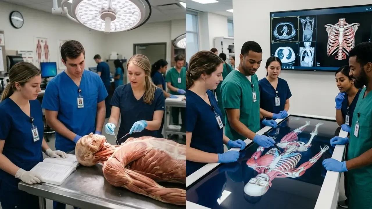
Cadaver Dissection vs. Virtual Anatomy Tools in PT Education
May 30, 2026Both cadaver dissection and virtual anatomy tools play crucial roles in physical therapy (PT) education. Each method has its strengths and limitations, and the best results often come from combining both approaches. Here's what you need to know:
- Cadaver Dissection: Offers hands-on experience with real cadavers, helping students understand natural anatomical variations and tactile details like muscle density. However, it involves high costs, limited accessibility, and exposure to chemicals.
- Virtual Anatomy Tools: Provide interactive 3D models, allow movement visualization, and are accessible anytime. Yet, they lack tactile feedback and can present idealized anatomy that doesn't reflect real patient variations.
Key Findings:
- Cadaver dissection benefits: Higher retention in identification skills, exposure to real anatomical variations.
- Virtual tools benefits: Better spatial visualization, integration with imaging like CT scans, and repeatability.
- Challenges: Cadavers are costly and logistically demanding; virtual tools lack tactile realism and can be expensive upfront.
Quick Comparison:
| Feature | Cadaver Dissection | Virtual Anatomy Tools |
|---|---|---|
| Tactile Feedback | Yes | No |
| Anatomical Variations | Yes | Limited |
| Accessibility | Restricted to labs | Anytime, anywhere |
| Cost | Ongoing (e.g., $1,200–$2,100 per donor) | High upfront (e.g., $200,000 for VDTs) |
| Dynamic Visualization | No | Yes |
Conclusion: Combining both methods bridges the gaps, offering a more complete learning experience. Cadaver dissection builds manual skills, while virtual tools enhance visual learning.
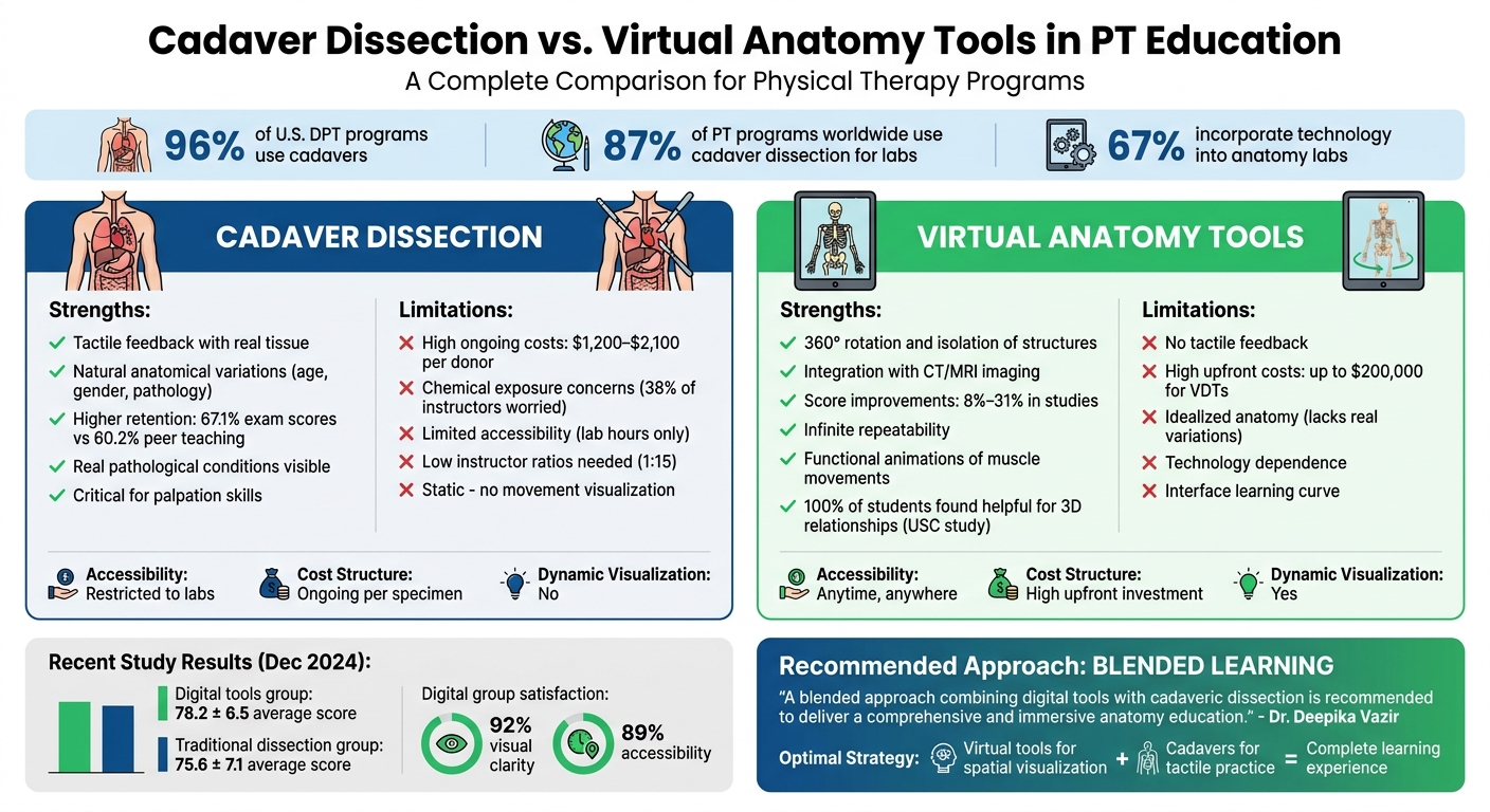
Cadaver Dissection vs Virtual Anatomy Tools: Complete Comparison for PT Education
Cadaver Dissection in Physical Therapy Education
Cadaver dissection plays a central role in teaching anatomy within physical therapy programs. A recent national survey found that over 90% of US DPT programs use donor-based dissection as part of their anatomy curriculum. This section dives into the advantages and challenges of using cadavers in physical therapy education.
Benefits of Cadaver Dissection
One of the biggest advantages of cadaver dissection is how it connects textbook learning to real-world clinical practice. Heather Henrichs from Nebraska Methodist College highlights this:
"Actually getting the chance to see the musculature of real cadavers in the lab creates an active, full-circle learning experience."
Cadavers reveal the natural variations in anatomy, such as differences based on gender, age, or health. They also show real-life pathological conditions like tumors, organ enlargement, or signs of past surgeries, helping students prepare for the unpredictable nature of clinical work.
The hands-on experience also has measurable benefits, with some comparative studies reporting higher practical exam performance for students who performed dissections than for those using certain alternative methods such as peer teaching. Andrew J. Saltarelli from Stanford University echoed this in his findings:
"Human cadaver laboratory offered a significant advantage over the multimedia simulation program on cadaver-based measures of identification and explanatory knowledge."
Dissection also sharpens students' ability to identify surface landmarks and understand spatial relationships - skills that are critical for palpation and manual therapy. However, this approach does come with its share of challenges.
Shocking Look Inside a Torn ACL (Cadaver Dissection Revealed)
Drawbacks of Cadaver Dissection
While cadaver dissection is effective, it isn’t without its challenges. One important concern is chemical exposure. Preservation chemicals like formaldehyde are a concern, with 38% of anatomy instructors reporting worries about their effects, and 11% believing their health is at risk.
There are also logistical hurdles. Dissections are expensive, require low instructor-to-student ratios (typically 1:15 or fewer), and are limited by reduced contact hours in competency-based education models. Adding to this, traditional "bell-ringer" practical exams - where students rotate through timed stations to identify anatomical structures - can be highly stressful. Many students report struggling to prepare for these exams and feeling rushed during the timed format compared to oral exams.
Virtual Anatomy Tools in Physical Therapy Education
Virtual tools are changing how anatomy is taught in physical therapy programs, offering alternatives to traditional cadaver dissection. This shift is especially important as anatomy contact hours are under pressure in many programs, and digital platforms are being used to help address reduced time and resource constraints.
Main Features of Virtual Anatomy Tools
Virtual dissection tables (VDTs) and 3D applications introduce features that go beyond what cadaver dissection can offer. These tools allow students to rotate models 360 degrees, isolate specific muscle groups, and examine anatomical structures from various angles - all without the logistical challenges of working with physical specimens.
Another key advantage is the integration of clinical imaging. VDTs can overlay CT scans and MRIs onto 3D models, helping students connect textbook concepts with diagnostic imaging they’ll encounter in practice. Some platforms even include functional animations, like demonstrating how muscles move during actions such as bending a knee or swallowing. These animations are particularly helpful for understanding movement, a cornerstone of physical therapy education.
The impact of these tools is supported by research. For instance, a study led by Dr. Deepika Vazir in June 2025 at a Government Medical College in India compared two groups of first-year medical students - one using 3D software and the Anatomage Table, and the other relying on traditional dissection. The digital group achieved an average post-test score of 78.2 ± 6.5, slightly higher than the 75.6 ± 7.1 scored by the traditional group. Additionally, 92% of the digital group reported satisfaction with visual clarity, and 89% appreciated the accessibility of the tools. Across various studies, the use of VDTs has been linked to academic score improvements ranging from 8% to 31%, with 86% of research noting better overall performance.
Another major benefit is repeatability. Unlike cadavers, which deteriorate with use, digital models can be reset instantly. This allows students to revisit complex structures as often as they need, free from time constraints or concerns about specimen availability. However, these tools are not without their limitations.
The BEST Way to Study Human Anatomy | An In Depth Look at the Kenhub Anatomy Platform
Drawbacks of Virtual Anatomy Tools
While virtual anatomy tools are highly interactive, they lack the tactile feedback crucial to physical therapy training. Physical therapists rely heavily on their sense of touch to assess muscle density, tendon tension, and other tissue characteristics - experiences that digital models simply cannot replicate.
Dependence on technology also poses challenges. In at least one recent study, most students reported enjoying digital tools, but those who studied with traditional atlases performed significantly better on specific anatomy tests, illustrating that digital resources do not always outperform conventional methods.
Cost is another hurdle. High-end virtual dissection tables can cost up to $200,000, making them a significant investment for educational programs. While cadaver preservation costs range from $1,200 to $2,100 per donor, the upfront expense of digital tools remains a barrier for many institutions.
Lastly, virtual tools typically present standardized versions of anatomy. Standardized models lack some of the individual variations – such as those caused by age, gender, pathology, or surgical history – that students encounter in real patients. This limitation can leave gaps in clinical readiness that only hands-on experience with cadavers can address.
Cadaver Dissection vs. Virtual Anatomy Tools
Learning Outcomes for Each Method
Both cadaver dissection and virtual anatomy tools bring distinct benefits to the table, shaping learning outcomes in different ways. Cadaver dissection is widely valued for improving identification and explanatory skills, offering students a hands-on experience with real human anatomy. On the other hand, virtual tools excel at simplifying spatial visualization, helping students grasp 3D structures without the mental strain of converting 2D textbook images into three-dimensional concepts.
"The human cadaver laboratory offered a significant advantage over the multimedia simulation program on cadaver-based measures of identification and explanatory knowledge." - Andrew J. Saltarelli, Office of the Vice Provost for Online Learning, Stanford University
Virtual tools also perform particularly well in some areas where traditional methods have limitations. At the University of Southern California, 100% of surveyed students found virtual anatomy apps helpful for understanding 3D relationships between structures, and every participant expressed a strong likelihood of using these tools in their future careers.
That said, the effectiveness of each method depends heavily on the user's experience and familiarity. Students who train with cadavers often outperform their peers in identifying and explaining structures. Meanwhile, virtual tools, despite their potential, can sometimes be hindered by interface challenges, especially for first-time users. The table below highlights the key differences between these two approaches.
Comparison Table: Cadaver Dissection vs. Virtual Anatomy Tools
| Feature | Cadaver Dissection | Virtual Anatomy Tools |
|---|---|---|
| Tactile Realism | High; offers an authentic feel of tissues and structures | None; relies on visual and auditory feedback only |
| Spatial Visualization | Requires mental conversion from 2D to 3D | High; provides interactive 3D models with rotation |
| Accessibility | Limited to lab hours and specific facilities | Flexible; accessible on tablets or mobile devices |
| Cost | High; involves facility upkeep and specimen sourcing | Lower; generally limited to software or app fees |
| Retention | Often strong for identification and explanatory knowledge in traditional curricula | Can match or exceed cadaver-based performance in some studies, though outcomes vary with design and user factors |
| Movement Visualization | Static; cannot show active muscle or joint movements | Dynamic; includes animated muscle actions and joint motion |
| Customization | None; limited to the donor's specific pathology | High; users can add features like fractures or inflammation |
As of a 2021 survey of U.S. Doctor of Physical Therapy programs, 90.5% of responding programs used cadaver dissection and 60.7% used computer-assisted technology in their anatomy teaching, reflecting a trend toward combining both methods. This data reflects a growing trend toward combining both methods, allowing students to benefit from the strengths of each approach.
Combining Both Methods for PT Education
Blended Learning for Better Results
Mixing cadaver dissection with virtual anatomy tools creates a balance that overcomes the shortcomings of each method. Cadavers provide real-world tactile experience and expose students to anatomical variations critical for building clinical confidence. Meanwhile, digital tools deliver interactive visualizations of muscle movements and joint mechanics - something static cadaver specimens can't replicate.
Recent studies highlight this synergy, with many reporting modest but meaningful improvements in exam scores when digital tools are combined with cadaver dissection compared with cadaver-only instruction. This underscores how combining the two approaches can enhance learning outcomes.
"A blended approach combining digital tools with cadaveric dissection is recommended to deliver a comprehensive and immersive anatomy education." - Dr. Deepika Vazir, Associate Professor, Government Medical College, Mulugu
This combined method also lightens the mental load for students. Instead of struggling to transform 2D textbook diagrams into 3D mental models, virtual tools offer ready-made, interactive visuals. These tools help students grasp spatial relationships, which they can then apply directly during cadaver labs. Here, they encounter real tissue variations and practice hands-on techniques essential for clinical practice. Together, these methods reinforce critical skills while making the learning process more manageable.
Institute of Human Anatomy's Role in Combined Learning
The Institute of Human Anatomy exemplifies this blended learning model by integrating cadaver-based education with digital resources. Their programs offer hands-on dissection opportunities with real human cadavers, providing students and professionals with the tactile experience needed to build clinical confidence. At the same time, they create educational videos, interactive courses, and digital guides, enabling learners to review and reinforce concepts outside the lab.
This dual approach ensures that both practical skills and dynamic visualization are developed simultaneously. For instance, in December 2024, 56 physical therapists enrolled in a postgraduate Sports Physiotherapy course at the University of Pisa participated in a one-day cadaver dissection course at the Institute of Human Anatomy, University of Bologna. All participants agreed that the hands-on experience significantly enhanced their clinical reasoning and core competencies. By combining physical lab sessions with accessible digital content, the Institute bridges the gap between theoretical learning and practical application, giving students the best of both worlds.
Conclusion
Cadaver dissection brings a hands-on, tactile experience that many educators view as important for clinical practice, reflected in survey data showing that about 90.5% of responding U.S. DPT programs include cadaver dissection in their anatomy teaching. On the other hand, virtual tools shine by offering interactive 3D visualization, which can ease cognitive load and help students grasp complex spatial relationships.
However, neither approach stands alone as fully sufficient. Research indicates that virtual dissection tables are linked to improved academic performance, with studies reporting score increases between 8% and 31%.
"Virtual dissection should supplement rather than replace traditional methods." - BMC Medical Education Systematic Review
The real advantage lies in combining these methods. Virtual tools can help students understand spatial relationships and anatomical structures dynamically, while cadaver dissection provides the tactile practice needed for real-world clinical scenarios. Together, they address the limitations of each method and offer a well-rounded educational experience.
Programs like those offered by the Institute of Human Anatomy are examples of programs that aim to use this blended approach, providing both cadaver-based training and digital resources. This combination equips students with the diverse skills they need to navigate the challenges of clinical practice effectively.
FAQs
Which anatomy method best prepares PT students for palpation and manual therapy?
Cadaver dissection is widely regarded as a key way for physical therapy (PT) students to build the skills they need for palpation and manual therapy. By working directly with real human tissue, students gain hands-on experience and a detailed understanding of anatomy. This includes recognizing different tissue textures and grasping how structures relate to one another spatially - both of which are crucial for developing manual therapy techniques.
While virtual tools can support visual learning, they lack the tactile feedback that only cadaver dissection can provide. The physical interaction with real tissue helps students develop the nuanced understanding necessary for effective patient care, something no simulation can fully replicate.
How can virtual anatomy tools be used without missing real anatomical variation?
Virtual anatomy tools tackle anatomical variation by combining case-based learning with multimodal approaches, helping students develop both clinical reasoning and spatial awareness. For instance, case-based virtual dissection introduces learners to a wide range of anatomical scenarios, giving them a deeper understanding of how anatomy can vary in real-life cases.
On top of that, working with multiple digital models that showcase different anatomical variations enhances students' ability to connect 2D images with 3D structures. This approach not only strengthens their grasp of anatomy but also complements traditional learning methods, making it easier to understand complex relationships within the human body.
What blended anatomy lab schedule works best in a DPT program?
The best blended anatomy lab schedule for a Doctor of Physical Therapy (DPT) program combines cadaver dissection with virtual anatomy tools. This mix offers a well-rounded learning experience by combining hands-on dissection with digital 3D applications and interactive demonstrations led by peers or faculty. By catering to different learning preferences, this approach keeps students engaged while balancing educational quality with cost considerations - an ideal setup for today’s PT education.

