
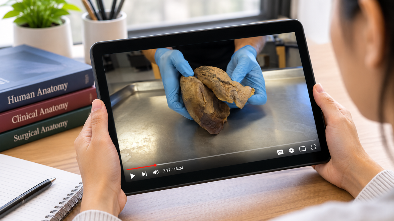
Teleanatomy: Exploring Anatomical Variations and Their Clinical Significance
Nov 03, 2026The human body is often presented in textbooks as a standardized blueprint. However, individual bodies differ in many ways. Anatomical structures can vary from person to person, sometimes in subtle ways and sometimes more noticeably. These differences, known as anatomical variations, and certain anatomical anomalies are more than interesting findings. They can have important implications for clinical anatomy, surgical anatomy, and patient safety. A newer framework in anatomy education, teleanatomy, described in a recent Clinical Anatomy article as “anatomy across distance,” is creating new ways to study this natural variation, especially for learners who can’t stand at a dissection table themselves.
Understanding these differences is essential for healthcare professionals. From interpreting diagnostic imaging to performing surgical procedures, recognizing that anatomical structures do not always follow textbook descriptions can help prevent misdiagnoses and complications. A thorough understanding of human anatomy is therefore an ongoing requirement in medical education and professional practice. Tools such as teleanatomy can further support this learning by giving students and practitioners access to detailed anatomical observations beyond the traditional classroom or dissection lab.
The Spectrum of Human Anatomy - More Than Just Textbooks
Anatomical variations are differences in the size, shape, position, or branching patterns of anatomical structures compared with the most commonly observed arrangement. They are generally not pathological and often represent normal differences among healthy individuals. In other words, the same anatomical structures may be arranged or shaped differently from one person to another without affecting normal function.
These differences occur throughout the body. Blood vessels, particularly arteries and veins, can have different branching patterns. For example, a renal artery may divide earlier than expected, or a person may have accessory renal arteries supplying the kidney. Nerves can also vary in their course, branching, or relationship to surrounding structures. Muscles may have additional muscle bellies, different insertion points, or variations in their size and shape. Some smaller muscles may also be absent in certain individuals. These findings are part of the natural diversity of human anatomy and are encountered regularly in anatomical study and clinical practice.
It is also important to distinguish healthy anatomical variations from anatomical anomalies. Anomalies generally refer to structural differences that are more significant and may be associated with altered development, impaired function, or clinical consequences. Recognizing this distinction helps healthcare professionals understand which findings represent normal variation and which may require further investigation. Both demonstrate why textbook illustrations should be treated as general references rather than exact representations of every individual.
Why Variations Matter: Impact on Clinical Practice
For healthcare professionals, recognizing anatomical variations is important for accurate diagnosis and appropriate treatment planning. Without this knowledge, a normal variation may be mistaken for a disease or abnormal growth, resulting in unnecessary tests or an incorrect diagnosis. On the other hand, an actual pathological condition may be overlooked when a practitioner is unfamiliar with the anatomical structures involved.
This is particularly relevant when interpreting diagnostic imaging. Radiologists may encounter anatomical variations that resemble tumors, cysts, or other abnormalities. An accessory spleen, a small extra nodule of splenic tissue, can resemble an enlarged lymph node or a tumor on imaging, including a mass in the tail of the pancreas. It can also enlarge after the main spleen is removed. Recognizing it for what it is can spare a patient an unnecessary biopsy or surgery. Similarly, an unusual blood vessel arrangement may be mistaken for a vascular abnormality. A strong understanding of clinical anatomy and common anatomical variations helps imaging specialists distinguish normal structures from conditions that require medical attention. This can prevent unnecessary procedures and support better patient safety.
Anatomical differences can also affect treatment decisions. A nerve that follows an unexpected course may require adjustments to an injection or other procedure to avoid injury. Likewise, an unusual blood supply to an organ may influence the approach to a biopsy, surgery, or stent placement. Effective patient care requires healthcare professionals to consider each patient's individual anatomy rather than assume that every body follows the same structural arrangement.
Precision is Paramount: Surgical Anatomy and Variations
Anatomical variations are particularly important in the operating room. Surgeons work within a complex, three-dimensional environment where the position and relationship of anatomical structures can directly affect the outcome of a procedure. An unexpected blood vessel, nerve, or organ position can make a routine operation more challenging and increase the risk of injury. This is why a thorough understanding of surgical anatomy is essential.
Consider a laparoscopic cholecystectomy, a common operation to remove the gallbladder. A 2026 meta-analysis pooling 27 studies of more than 9,600 patients found conventional cystic artery anatomy in about 86% of cases observed during surgery, meaning roughly one in seven patients had some variation. Among the most common were a duplicate cystic artery (about 7%) and a “caterpillar hump” (about 6%). A caterpillar hump is a variation of the right hepatic artery, a major vessel supplying the liver: instead of running straight, it loops into the area surgeons dissect, often leaving a very short cystic artery beside it. Variations like these can make it harder to identify structures correctly and may increase the risk of injury to blood vessels or bile ducts, which is why surgeons are trained to confirm what each structure is before dividing it. Similarly, in orthopedic surgery, variations in the pathways of nerves around a joint can increase the risk of nerve injury during implant placement or other procedures.
Preoperative planning is therefore essential. Diagnostic imaging, including CT and MRI scans, can help surgical teams identify relevant anatomical variations and anatomical anomalies before an operation. With this information, surgeons can anticipate potential challenges, adjust their approach, and prepare for unexpected findings. However, imaging is only one part of the process. Surgeons must also be able to recognize and respond to the structures they encounter during surgery. A strong foundation in human anatomy and surgical anatomy helps them make informed decisions and reduce risks to patient safety.
The Cornerstone of Understanding: Cadaveric Dissection and Medical Education
How do healthcare professionals develop a practical understanding of anatomical variations? Textbooks, illustrations, and 3D models provide valuable foundational knowledge, but cadaveric dissection remains a central part of anatomy education. Working with human cadavers gives students the opportunity to examine anatomical structures in three dimensions, observe their relationships, and identify differences that may not be apparent in illustrations or digital models.
At the same time, medical education continues to expand beyond the traditional dissection lab. Teleanatomy describes anatomy studied across distance: anatomical findings captured in one place, such as a cadaver lab, and studied or applied somewhere else. Rather than being tied to a specific technology, the framework focuses on how anatomical information is shared remotely and whether it stays accurate along the way. A filmed dissection of a real donor body, watched from another city, is one example. This allows students and healthcare professionals to observe anatomical structures and variations from different specimens, even when they cannot attend an anatomy lab in person.
At the Institute of Human Anatomy (IOHA), we use real human cadaver dissections to teach anatomy through educational videos, online courses, and live-streamed workshops. We share our content with millions of learners through YouTube and our online learning platform. Through our cadaveric dissections, we give learners the opportunity to observe real human anatomical structures, including variations that may not appear in standard anatomy illustrations or textbooks. In the video below, Jonathan Bennion, PA-C, shows a right lung with only two lobes instead of the usual three and explains why this variation doesn't necessarily affect how the lung works.
5 Craziest Things I've Found In Dead Bodies
Footage like this lets learners see actual anatomical structures, follow an expert demonstration, and rewatch specific areas as often as they need, building a sense of how much real bodies differ from one another.
In a traditional cadaver lab, students can encounter anatomical differences firsthand. A textbook may show one commonly observed branching pattern of an artery, while examination of multiple cadavers can reveal other arrangements. Seeing these differences helps students understand that textbook illustrations represent typical anatomy, not necessarily the exact anatomy of every patient. It also encourages them to approach clinical situations with greater awareness rather than relying on a single expected arrangement.
Teleanatomy complements hands-on learning by making recorded dissections available for repeated observation. Students can review complex structures, compare anatomical findings, and study regions that may not have been covered in depth during their in-person sessions. This wider access can strengthen exposure to anatomical diversity and support more comprehensive medical education.
Cadaveric dissection also develops spatial reasoning and provides direct experience with the relationships and textures of human tissues. These aspects of learning are difficult to reproduce through textbooks or digital resources alone. It remains an important foundation for understanding clinical anatomy and surgical anatomy, while teleanatomy and other digital learning tools provide additional ways to study and review the body's anatomical structures.
Beyond the Dissection Lab: Diagnostic Imaging and Advanced Techniques
Although cadaveric dissection is essential for studying anatomy, healthcare professionals must also understand how anatomical structures appear in living patients. Advanced diagnostic imaging techniques, including computed tomography (CT), magnetic resonance imaging (MRI), and ultrasound, allow clinicians to examine internal structures and identify individual anatomical differences. These tools are especially useful when assessing a patient's anatomy before surgery or another medical procedure.
Combining cadaveric study, teleanatomy, and modern imaging helps students connect what they see in dissection with how the same structures appear on CT, MRI, and ultrasound. Research suggests this kind of integration can improve learning, though results vary between studies. Either way, it builds a skill clinicians rely on every day: recognizing the same structure in very different views.
For example, a student who has studied the branching patterns of arteries during dissection can apply that knowledge when examining vascular structures on a CT scan. Understanding how normal variations appear in both cadavers and living patients can help future healthcare professionals interpret diagnostic imaging more accurately and recognize differences before they become unexpected findings during a procedure. These learning approaches support the development of practitioners who can apply their knowledge of clinical anatomy and surgical anatomy in real clinical settings.
Fostering a Culture of Awareness for Enhanced Patient Safety
Understanding anatomical variations is not simply an academic requirement. It is an important part of providing safe and effective patient care. Healthcare institutions and medical education programs should emphasize the recognition of anatomical differences as a core professional skill. This includes:
- Robust Anatomy Curricula: Providing sufficient opportunities for students to study anatomical variations through cadaveric dissection, teleanatomy, and other anatomy learning resources.
- Continuing Professional Development: Encouraging healthcare professionals, particularly surgeons and radiologists, to stay informed about anatomical variants and advances in diagnostic imaging.
- Critical Thinking: Teaching clinicians to consider possible anatomical variations when interpreting imaging results, planning procedures, and evaluating patient conditions.
- Interdisciplinary Collaboration: Encouraging communication among radiologists, surgeons, and other healthcare specialists when anatomical differences may affect diagnosis or treatment.
Making anatomical awareness a consistent part of healthcare education and practice helps professionals make better-informed decisions, reduce avoidable risks, and provide more appropriate care for individual patients.
Frequently Asked Questions about Teleanatomy
What is teleanatomy?
Teleanatomy is anatomy across distance. A 2026 article in Clinical Anatomy proposed it as a conceptual framework for situations where anatomical information is captured in one place and studied, taught, or applied somewhere else. That can include a live-streamed or filmed cadaver dissection, remote teaching with 3D models or virtual reality, or anatomical information shared between clinicians in different locations. What defines it is not the technology used, but the distance, and whether the anatomy stays accurate and reliable as it travels.
How does teleanatomy enhance medical education?
Teleanatomy gives students and healthcare professionals flexible access to anatomical learning materials. They can revisit complex dissections, review anatomical structures, and observe variations across different specimens. It also makes anatomy resources more accessible to learners who do not have regular access to a physical dissection lab.
Can teleanatomy replace traditional cadaveric dissection?
Teleanatomy is a valuable addition to anatomy education, but it does not fully replace traditional cadaveric dissection. Working directly with human cadavers provides hands-on experience, helps develop spatial reasoning, and allows students to examine tissue structures and relationships firsthand. Teleanatomy supports this learning by providing additional demonstrations, repeated viewing, and broader exposure to anatomical differences.
How does teleanatomy help in understanding anatomical variations?
Recorded and live-streamed dissections let learners see the same structure in many different bodies, which is hard to do in a typical lab course where students may work with only one or two donors. Seeing differences in size, shape, position, and branching patterns across specimens builds the habit of expecting variation rather than assuming textbook anatomy. Studies of remote cadaver-based teaching show it can support solid learning outcomes, but research specifically on recognizing variations is still limited, which is why we see teleanatomy as a complement to hands-on dissection, not a replacement.
Who benefits from teleanatomy?
A range of learners and professionals can benefit from teleanatomy, including:
- Medical and healthcare students: For foundational anatomy learning, coursework, and revision.
- Practicing clinicians: For reviewing anatomical structures and refreshing their knowledge of clinical anatomy and surgical anatomy.
- Remote learners: Those who have limited access to physical anatomy laboratories.
- Researchers: For reviewing anatomical structures and studying variations across specimens.
- Educators: For demonstrating anatomical structures and explaining complex concepts during instruction.
Conclusion
A thorough understanding of human anatomy is essential for safe and effective healthcare practice. Anatomical variations are a normal part of human diversity and can have important implications for clinical anatomy, surgical anatomy, and patient safety. Through cadaveric dissection, advanced diagnostic imaging, and digital learning approaches such as teleanatomy, students and healthcare professionals can develop a broader understanding of how anatomical structures differ between individuals. These learning methods help prepare future practitioners to recognize anatomical differences, interpret clinical findings, and approach procedures with greater awareness of each patient's anatomy.
Want to see anatomical variations for yourself? Explore our human anatomy videos on the Institute of Human Anatomy YouTube channel, where we use real human cadavers and visual demonstrations to explain anatomical structures and variations. You can also join one of our live-stream Anatomy & Physiology workshops to study real human anatomy from wherever you are.
Sources
- Iwanaga J, Tubbs RS. “Teleanatomy: A Conceptual Framework for Anatomy Across Distance.” Clinical Anatomy, 2026. https://doi.org/10.1002/ca.70247
- Triantafyllou G, et al. “Intraoperative Variations of Cystic Artery and Duct in Laparoscopic Cholecystectomy: A Systematic Review With Meta-Analysis.” ANZ Journal of Surgery, 2026. https://doi.org/10.1111/ans.70651
- Alexander M, Montalbano MJ, Loukas M. “Learning Anatomy With Radiology: A Systematic Review.” Clinical Anatomy, 2026. https://doi.org/10.1002/ca.70048
- Hunter S, Halliday NL, Reeder S, Garman L, Moon MB. “Clinical Anatomy Remote Learning: A Mixed Methods Analysis of Learning Outcomes and Student Perceptions.” Anatomical Sciences Education, 2026. https://doi.org/10.1002/ase.70165

