
What Is HuBMAP? Exploring the Healthy Human Tissue Atlas
Nov 04, 2026The intricacies of the human body have captivated scientists, educators, and the curious public for centuries. Understanding how our bodies work, from the largest organ systems down to the tiniest cells, is fundamental to both medical advancement and personal well-being. But what if we could access an incredibly detailed reference of healthy human tissues? What if this baseline could help researchers better understand the changes associated with disease?
This vision is rapidly becoming a reality. In a groundbreaking development, researchers and data scientists from numerous institutions, including Harvard Medical School, have collaborated on an ambitious, federally funded initiative known as the Human BioMolecular Atlas Program (HuBMAP). This monumental effort has culminated in the launch of the HuBMAP Data Portal – a free, open-access online resource that offers an extensive human anatomy atlas of healthy human tissue available in a single platform. Described in a recent article in Nature Communications, this project represents an eight-year endeavor involving over 600 consortium members from more than 60 institutions, supported by the National Institutes of Health (NIH) Common Fund.
This cutting-edge digital atlas isn't just a collection of pretty pictures; it’s a robust, dynamic resource providing access to over 9,000 datasets from more than 500 donors, spanning dozens of organs and tissues across 29 organ classes. Its datasets include data at single-cell and near-single-cell resolution, offering detailed views of healthy human tissues. For the medical education community, researchers, and even those with a general interest in human anatomy, this atlas offers a valuable resource for advancing our understanding of health and disease by providing a detailed reference map of healthy human tissue.
The Power of a Healthy Baseline: Connecting the Cadaver Lab to the Digital Frontier
For generations, the cornerstone of medical education and anatomical understanding has been the hands-on experience of cadaveric dissection. In labs around the world, students and researchers meticulously explore the intricate network of muscles, nerves, blood vessels, and organ systems that constitute the human form. This direct interaction with a real human body provides a distinctive appreciation for the complexity and interconnectedness of our biology. It is here, on the dissection table, that the foundational understanding of what a "normal" or "healthy" body looks often begins.
At its core, cadaveric dissection provides a macroscopic anatomical baseline. It allows us to grasp the relative positioning of organs, the branching patterns of arteries, and the precise pathways of nerves. Without this fundamental understanding of the healthy state, identifying and comprehending pathological conditions – diseases, injuries, or anomalies – would be a far more challenging, if not impossible, task. The question becomes: how do we recognize what's wrong if we don't profoundly understand what's right?
From Gross Anatomy to the Microscopic Realm
While the cadaver lab excels at revealing gross anatomy – the structures visible to the naked eye – modern science demands a deeper dive. Disease often manifests not just at the organ level, but at the tissue and cellular levels. This is where HuBMAP extends and amplifies the concept of an anatomical baseline.
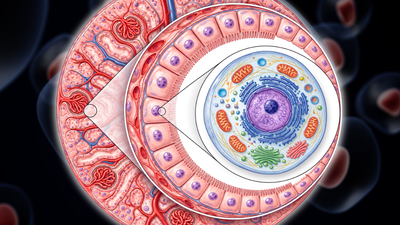
Imagine moving from the panoramic view of an entire organ in a cadaver, to zooming in on a specific tissue type within that organ, and then further still, to examine the individual cells that compose that tissue. HuBMAP provides precisely this multi-scale perspective, offering an unprecedented look at healthy human tissue down to its fundamental building blocks.
For instance, a medical student might observe the healthy morphology of a kidney during a cadaveric dissection. HuBMAP allows them (or an AI algorithm) to then access detailed healthy body data, showing the precise cellular composition of a healthy kidney, the gene expression patterns within those cells, and how different cell types interact across the organ systems. This integration of macroscopic and microscopic healthy body data is crucial. A tumor, for example, might be visible as a mass in gross anatomy, but its true nature – its cell biology, its genetic mutations, and its impact on surrounding healthy human tissue – can be better understood through comparison with detailed references of healthy human tissue.
This comprehensive approach is designed to help researchers:
- Identify subtle deviations: With healthy tissue as a point of comparison, HuBMAP data can help researchers investigate subtle cellular and molecular changes that may be associated with early disease.
- Understand disease progression: Researchers can track how diseases alter healthy human tissue over time, from initial cellular changes to macroscopic pathology.
- Support therapy development: Detailed data on healthy cellular function can help inform research into therapies that target disease-related changes while considering their effects on healthy cells.
- Support AI research: The extensive datasets provide valuable resources for training artificial intelligence models and benchmarking computational tools used in biological research.
The HuBMAP Data Portal, with its digital human anatomy atlas, therefore acts as a critical bridge. It takes the foundational knowledge gained from traditional medical education and cadaveric dissection and elevates it to a new dimension, providing a granular reference for healthy human tissue across multiple organ classes. This gives federally funded research, such as that conducted at Harvard Medical School and other NIH-supported institutions, a shared baseline for comparison when studying how to prevent and treat disease.
The Legacy of Generosity: The Donor Story
None of this profound understanding – neither the hands-on insights from the cadaver lab nor the intricate digital maps of HuBMAP – would be possible without an extraordinary act of human generosity: body donation.
In the cadaver labs, families make the deeply personal decision to donate a loved one's body to science and medical education. This selfless act provides an invaluable gift to countless students and researchers, offering them the unique opportunity to learn from a real human form. It is a legacy that directly shapes the next generation of medical professionals and contributes to diagnostic and surgical advancements.
Similarly, the HuBMAP project stands as a testament to the generosity of over 500 healthy individuals who donated their tissues for this ambitious undertaking. These donors, driven by a desire to contribute to the greater good, provided the healthy human tissue samples that form the very foundation of this comprehensive human anatomy atlas. Each sample, meticulously collected and analyzed, contributes to the vast pool of healthy body data that will serve as a reference point for researchers globally.
The act of body donation, whether for cadaveric dissection or for tissue sampling in projects like HuBMAP, underscores a powerful human truth: our bodies, even after life, can continue to teach, heal, and inspire. These donors become silent, yet profound, contributors to scientific progress, directly impacting our collective understanding of human health. Their generosity contributes to the anatomical reference and research resources that can support future medical discoveries.
This is What Happens When You Die
A New Era for Medical Science and Education
The launch of the HuBMAP Data Portal marks a significant milestone in biomedical research, with potential value for medical science and education. By providing an open-access atlas of healthy human tissue, it gives researchers and clinicians a valuable resource for understanding the healthy body and, by extension, the changes associated with disease.
The macroscopic insights gained through cadaveric dissection and the microscopic details captured in HuBMAP can complement each other in building a more complete understanding of the human anatomical baseline. This combined approach, spanning from the visible structures in a cadaver lab down to the intricate cell biology of every organ system, is intended to support a multi-scale view of human health. It is designed to help researchers identify disease and develop treatments, and ultimately, improve human lives.
This endeavor brings together dedicated scientists from more than 60 institutions, including Harvard Medical School, with support from the NIH Common Fund and contributions from human tissue donors. Their collaborative work provides an extensive reference for studying healthy human tissues.
More than just an atlas, it represents a valuable resource for future discoveries, a testament to collaboration, and an important step toward a more comprehensive understanding of the human body.
Sources
-
Turner ML, Smits TC, Liaw TS, et al. (2026). HuBMAP Data Portal: a resource for multimodal spatial and single-cell data of healthy human tissues. Nature Communications, 17, Article 11125.

