
Neurodegenerative Diseases Linked to Vagus Nerve
Jun 26, 2026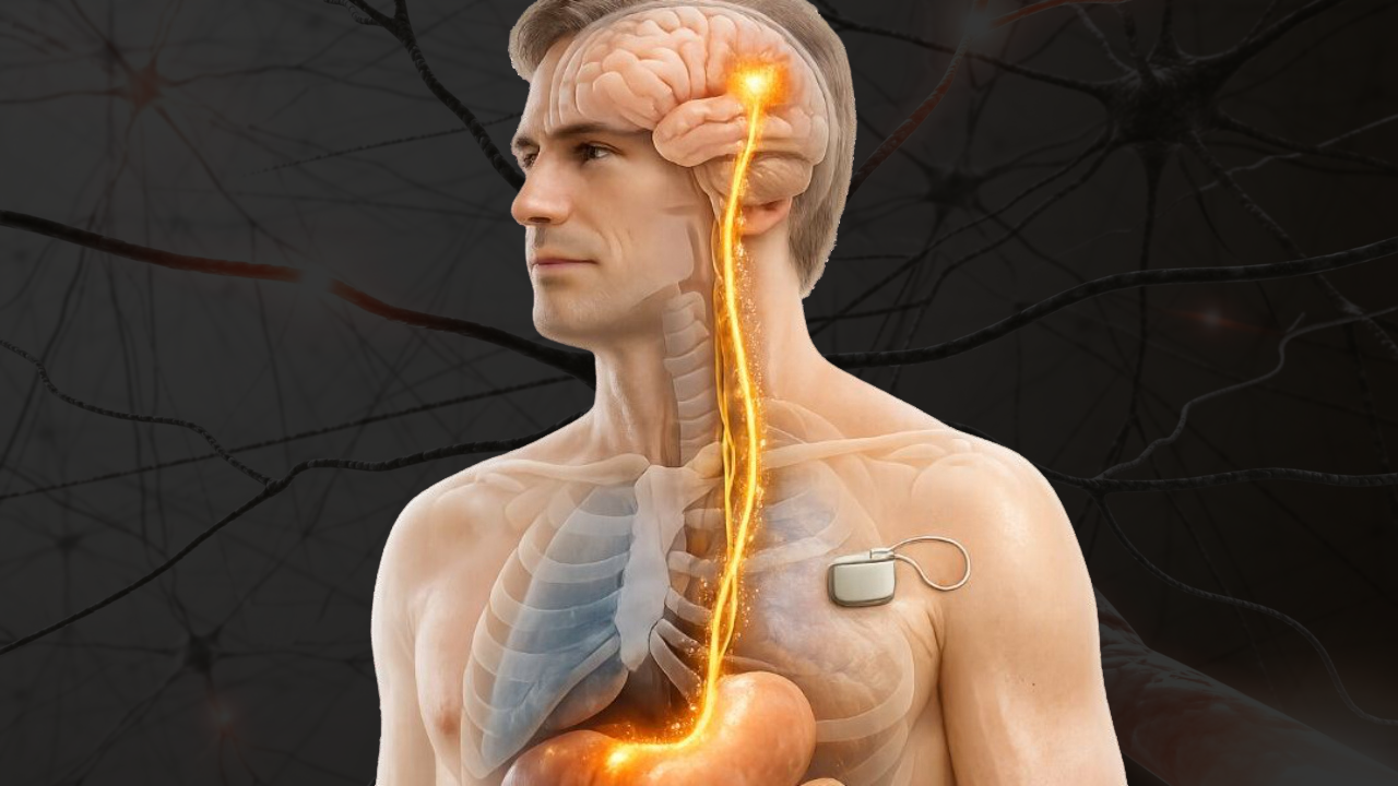
The vagus nerve, a critical part of the parasympathetic nervous system, has emerged as a key player in neurodegenerative diseases like Parkinson’s and Alzheimer’s. Experimental studies show that misfolded α-synuclein in Parkinson’s can propagate from the gut to the brain via the vagus nerve in animal models, and similar gut–brain routes are being explored for Alzheimer’s disease, although these mechanisms remain under active investigation in humans. This connection explains early symptoms like digestive issues and highlights the nerve's role in disease progression through inflammation and protein aggregation.
Key points:
- Parkinson's Disease: Misfolded α-synuclein can spread from the gut to the brainstem in some experimental models and may do so in a subset of patients, contributing - to motor and autonomic dysfunction.
- Alzheimer's Disease: Disturbances in brainstem and basal forebrain circuits, including those involving cholinergic and noradrenergic neurons that interact with vagal pathways, contribute to reduced acetylcholine signalling and cognitive decline.
- Vagus Nerve Stimulation (VNS): Emerging as a potential therapy to reduce inflammation, improve neurotransmitter activity, and may slow disease progression, although this remains unproven in humans.
The vagus nerve's role in the gut-brain axis and its potential for therapeutic intervention make it a focal point in neurodegenerative research and education.
How Neurodegenerative Diseases Affect the Vagus Nerve
Protein Aggregation in Vagal Pathways
The vagus nerve plays a crucial role in how neurodegenerative diseases progress, particularly through the buildup of pathological proteins and related inflammatory responses.
In Parkinson's disease, the misfolding and accumulation of α-synuclein (aSyn) occur in the dorsal motor nucleus of the vagus (DMV) - a brainstem region responsible for the nerve's motor functions.
"The dorsal motor nucleus of the vagus nerve (DMnX) is a primary site of pathological α-synuclein deposition and may play a key role in the spreading of α-synuclein lesions within and outside the CNS." - Ruth E. Musgrove et al.
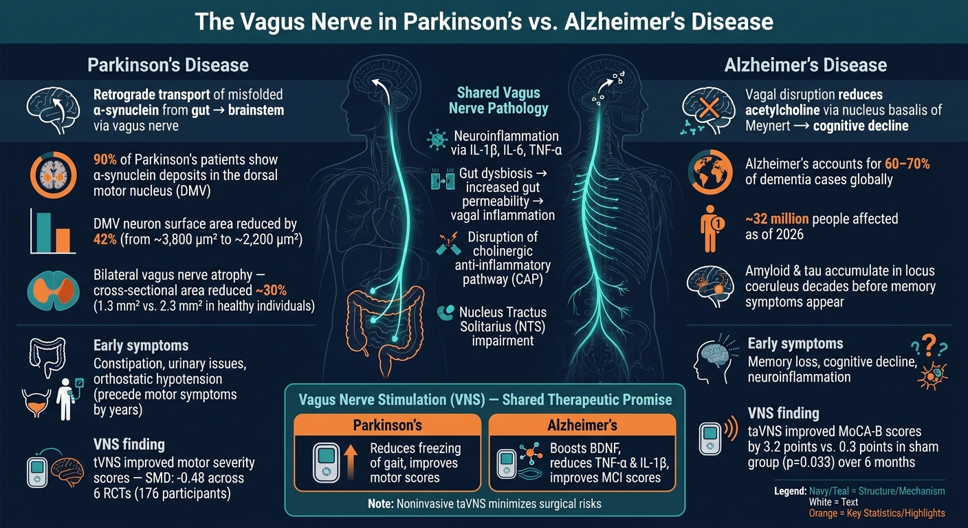
Neuropathological studies indicate that many Parkinson’s patients show α-synuclein deposits in the dorsal motor nucleus of the vagus, which is considered an early and vulnerable site of pathology. In experimental models, excessive aSyn reduces the DMV neuron surface area by 42% (from ~3,800 μm² to ~2,200 μm²). Experimental studies suggest that α-synuclein-related changes in ion channel function can slow pacemaker activity in DMV neurons, potentially weakening vagal parasympathetic output, but these mechanisms are still being clarified.
Interestingly, enteroendocrine cells (EECs) in the gut lining - cells with neuron-like structures called "neuropods" - also express aSyn. These cells connect directly to vagal sensory neurons, suggesting they might be an early site for protein misfolding before it spreads to the brain.
This protein buildup sets off inflammatory responses, further damaging vagal function.
Neuroinflammation and Autonomic Dysfunction
The vagus nerve's impairment is worsened by systemic inflammation within the gut–brain axis. Activated microglia and astrocytes release pro-inflammatory cytokines like IL-1β, IL-6, and TNF-α, which interfere with the Nucleus Tractus Solitarius (NTS) - the brain's hub for processing vagal sensory signals.
"The NTS serves as a cytokine detector within the CNS, and the local synthesis of inflammatory mediators directly influences neuronal functionality in the NTS." - Frontiers in Aging Neuroscience
The effects of this inflammation are far-reaching. Reduced DMV activity leads to a decline in parasympathetic vagal tone, impairing gut motility. This explains why constipation and slowed digestion often precede motor symptoms in Parkinson's disease by several years. Oxidative stress adds to the problem by creating nitrated forms of aSyn, which are even more prone to spreading between cells, accelerating degeneration in the DMV.
The Gut–Brain Axis and Vagus Nerve Communication
The vagus nerve serves as the main physical link between the gut and brain. When gut bacteria become imbalanced (gut dysbiosis), it can trigger intestinal inflammation and increase gut wall permeability. This allows inflammatory signals to reach the vagus nerve's enteric connections, enabling the retrograde transport of aSyn toward the brain.
The structural damage to the vagus nerve is stark. Parkinson's patients exhibit bilateral atrophy of the vagus nerve, with studies reporting a reduced cross-sectional area compared to healthy individuals of the same age. As Professor of Neurology Uwe Walter explains, "PD is associated with a bilateral atrophy of the vagus nerve but not of the spinal accessory or the phrenic nerves". This points to a disease process that specifically targets vagal tissue. Adding weight to the gut-to-brain hypothesis, some studies suggest that individuals who undergo complete truncal vagotomy have a lower risk of developing Parkinson's disease, supporting the idea that, in some cases, the vagus nerve may serve as a route for disease-related pathology.
Parkinson’s Disease May Start in the Gut
Neurodegenerative Diseases That Affect the Vagus Nerve
Vagus Nerve in Parkinson's vs. Alzheimer's Disease: Key Mechanisms & Data
Parkinson's Disease and Synucleinopathies
Parkinson's disease provides a striking example of how the vagus nerve is involved in neurodegeneration. In this condition, misfolded α-synuclein proteins travel backward along the vagus nerve from the gut to the brainstem, specifically targeting the dorsal motor nucleus (DMV). In cases of "body-first" Parkinson's, this retrograde movement of α-synuclein precedes the onset of motor symptoms. Early signs often include autonomic issues like constipation, urinary problems, and orthostatic hypotension. On the other hand, "brain-first" Parkinson's originates in the brain or olfactory system and spreads outward, typically bypassing these early gut-related symptoms.
"The vagus nerve has been repeatedly suggested to represent one major route of disease progression in Parkinson's disease (PD), with an active retrograde transport of α-synuclein originating in the enteric nervous system ascending the vagus nerve and eventually reaching the dorsal motor nucleus of the vagus (dmX)." - Uwe Walter, Professor of Neurology, University of Rostock
The damage isn't limited to neurons. Deposits of phosphorylated α-synuclein (p-α-syn) in the Schwann cells of the vagus nerve trigger inflammation through Toll-like receptor 2 (TLR2). This inflammatory response leads to Schwann cell death and demyelination, further impairing the nerve's function.
"Our results support the perspective that p-α-syn interacts with TLR2 induced SCs [Schwann cells] damage and is involved in PD AutD [autonomic dysfunction]." - Cell Death Discovery
These findings highlight the vagus nerve's central role in Parkinson's progression, where it serves as a direct pathway for disease-related proteins. In contrast, Alzheimer's disease shows a more indirect but equally critical connection to vagal dysfunction.
Alzheimer's Disease and Dementias
While Parkinson's involves direct protein propagation via the vagus nerve, Alzheimer's disease impacts the nerve through disruptions in the brain's cholinergic system.
Alzheimer's, which accounts for 60–70% of dementia cases globally, affects tens of millions of people worldwide, with prevalence projected to rise substantially over coming decades. The vagus nerve plays a less visible but still crucial role in the disease's progression.
A key link lies in the cholinergic system of the basal forebrain, including the nucleus basalis of Meynert, which is the primary source of cortical acetylcholine, a neurotransmitter essential for memory and learning 9. Brainstem nuclei that receive vagal input, together with basal forebrain and locus coeruleus projections, form interconnected networks that modulate cholinergic and noradrenergic signalling. In Alzheimer’s disease, degeneration within these networks contributes to reduced acetylcholine availability and cognitive decline.
Another early indicator involves the locus coeruleus (LC), a brainstem region that receives input from the vagus nerve. Research shows that amyloid and tau proteins start accumulating in the LC decades before memory symptoms appear.
"The accumulation of amyloid and tau in the LC is an early pathological feature preceding clinically detectable memory impairment by decades." - Ruth Narramore, Sheffield Teaching Hospitals NHS Foundation Trust
The vagus nerve also plays a role in regulating inflammation through the cholinergic anti-inflammatory pathway (CAP). When these regulatory pathways are impaired, the brain's immune cells - microglia and astrocytes - become overactive. This leads to the release of inflammatory cytokines, which hasten neural damage.
Alzheimers vs Dementia
Below is a breakdown of how vagal dysfunction contributes to Alzheimer's pathology:
| Mechanism | What Goes Wrong | Result |
|---|---|---|
| Cholinergic pathway disruption | Reduced acetylcholine synthesis | Memory loss, cognitive decline |
| Neuroinflammation | Overactive microglia; IL-1β, TNF-α release | Accelerated neural damage |
| Protein aggregation | Spread of Aβ plaques and tau tangles | Progressive neurodegeneration |
| HPA axis dysregulation | Increased cortisol due to vagal dysfunction | Damage to hippocampal neurons |
This evidence underscores the vagus nerve's involvement in both motor and cognitive declines, making it a critical focus in understanding neurodegenerative diseases.
Vagus Nerve Stimulation as a Potential Treatment
Vagus nerve stimulation (VNS) involves sending electrical impulses along the vagus nerve, either through surgical implants or noninvasive methods targeting the ear or neck. This stimulation can encourage neurotransmitter release and reduce inflammation. Below, we’ll dive into how VNS is showing promise in treating Parkinson’s disease and Alzheimer’s, along with the challenges researchers face.
VNS in Parkinson’s Disease
For Parkinson’s disease, VNS appears to enhance motor function by influencing dopamine production, reducing inflammation, and may reduce α‑synuclein aggregation in preclinical models, a protein linked to the disease's progression. A meta-analysis conducted in 2025, which reviewed six randomized controlled trials (RCTs) with 176 participants, found that transcutaneous VNS (tVNS) significantly improved motor severity scores and gait during "on-medication" phases. The standardized mean difference (SMD) was -0.48 for both measures. Another study by Mondal and colleagues highlighted that tVNS could meaningfully reduce freezing of gait, a particularly disabling symptom of Parkinson’s.
"The capacity of VNS to regulate LC activity, mitigate inflammation, and modulate ANS function demonstrates its considerable therapeutic promise in addressing the complexities of PD pathology." - npj Parkinson’s Disease
VNS in Alzheimer’s and Cognitive Disorders
Preclinical and early clinical research suggests that VNS can influence cholinergic signaling, reduce inflammatory cytokines like TNF‑α and IL‑1β, and may increase brain-derived neurotrophic factor (BDNF), which aids neuron survival. A 2022 double-blind clinical trial by Wang et al. assessed transcutaneous auricular VNS (taVNS) in patients with mild cognitive impairment (MCI). Over six months, taVNS improved MoCA-B scores by 3.2 points, compared to just 0.3 points in the sham group - a statistically significant difference (p = 0.033).
"VNS provides a potential therapy that can protect against the progression of Alzheimer’s disease at its earliest stage." - Ruth Narramore, Sheffield Teaching Hospitals NHS Foundation Trust
Remaining Challenges and Future Research
While early results are promising, there are hurdles to overcome. Many human trials have small sample sizes and inconsistent stimulation protocols, making it difficult to draw firm conclusions. Researchers suggest that future studies should include objective biomarkers - like VSEP, fMRI, or EEG - to ensure accurate measurement of treatment effects and to refine stimulation parameters and lateralization strategies.
"Clinical results remain inconsistent due to variability in treatment duration, outcome measures, and reliance on subjective assessments." - Current Alzheimer Research
Noninvasive approaches, especially taVNS, are gaining traction as they reduce risks like infection, hoarseness, and swallowing difficulties associated with surgical implants. These methods also come with minimal side effects, such as mild skin irritation. As technology advances and study designs improve, VNS might move from experimental status to a widely accepted therapy for neurodegenerative diseases.
Teaching Vagus Nerve Pathology Through Cadaver-Based Anatomy
Studying the Vagus Nerve Using Human Cadavers
Textbooks can outline the vagus nerve's anatomy, but nothing compares to physically tracing its path in a cadaver. Dissection allows students to follow the nerve from the brainstem through the chest and abdomen, linking its structure to clinical symptoms seen in neurodegenerative diseases.
One of the standout benefits of cadaveric study is the ability to identify key brainstem nuclei, such as the dorsal motor nucleus of the vagus (DMV), the Nucleus Tractus Solitarius (NTS), and the Locus Coeruleus (LC). These areas are some of the first to show α-synuclein pathology in Parkinson’s disease, with the LC being affected as early as Braak Stage 2. Observing these nuclei firsthand helps students grasp why autonomic issues, like persistent constipation, often precede motor symptoms by years. Additionally, cadaver studies highlight the importance of the nerve's fiber composition. For example, the selective loss of unmyelinated viscero-afferent and viscero-efferent fibers - while sparing thicker myelinated motor fibers - explains the autonomic dysfunctions commonly seen in Parkinson’s. This hands-on exploration bridges the gap between theoretical concepts and clinical observations.
Using Pathological Specimens to Show Disease Progression
While cadaver dissections provide foundational anatomical insights, examining pathological specimens offers a deeper look into disease progression. Unlike healthy cadavers, these specimens reveal the physical effects of neurodegeneration, giving students a tangible understanding of conditions like Parkinson’s.
The data is striking. In patients with Parkinson’s disease, ultrasound studies report that the vagus nerve cross‑sectional area is modestly but significantly smaller than in healthy individuals on both sides. Meta‑analyses indicate a bilateral reduction in CSA consistent with vagus nerve atrophy, although the absolute differences are relatively small.
"PD is associated with a bilateral atrophy of the vagus nerve but not of the spinal accessory or the phrenic nerves." - Uwe Walter, Department of Neurology, University of Rostock
Pathological specimens also allow students to trace the spread of α-synuclein, starting in the enteric nervous system, moving through the DMV, and eventually reaching higher brainstem regions. This hands-on experience reinforces the mechanisms of protein propagation covered in earlier discussions.
How the Institute of Human Anatomy Supports This Learning
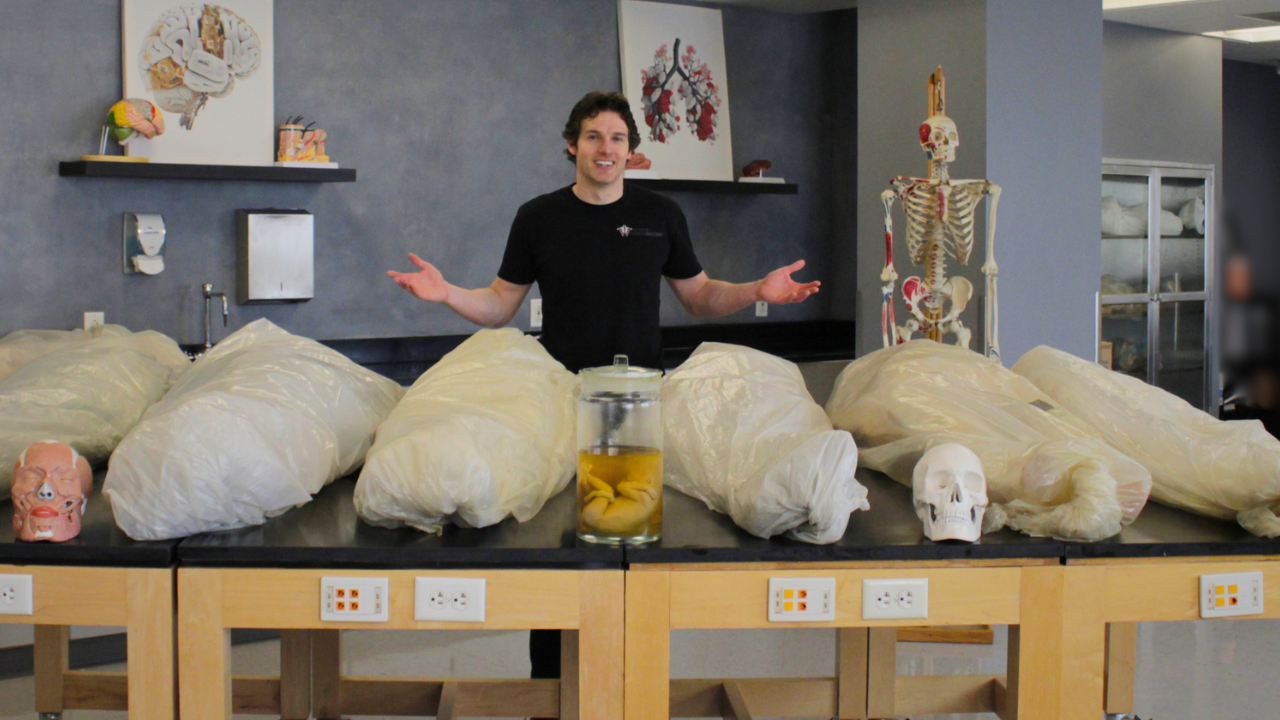
The Institute of Human Anatomy plays a pivotal role in making these learning experiences accessible. Based in Salt Lake City, Utah, the Institute focuses on teaching human anatomy through cadaver-based methods, emphasizing both anatomical accuracy and clinical relevance.
The Institute offers a range of resources, including interactive courses, digital study guides, eBooks, and comprehensive bundles covering all major body systems. For students seeking extra help, an AI-powered Interactive Anatomy & Physiology Buddy is available 24/7 to answer questions and provide tailored study support. With over 24 million followers and subscribers and more than 2 billion video views, the Institute has become one of the most widely accessible resources for cadaver-based education.
"Using real human cadavers to teach as many people as possible about the one thing we all have in common - our bodies." - Institute of Human Anatomy
Conclusion: The Vagus Nerve in Neurodegenerative Research and Education
The vagus nerve holds a pivotal role in neurodegenerative research. Acting as a pathway for the retrograde transport of harmful proteins like α‑synuclein, it contributes to neuroinflammation and facilitates communication along the gut–brain axis. These mechanisms provide valuable insights into the origins and progression of conditions such as Parkinson's and Alzheimer's.
One of the most promising areas of exploration is vagus nerve stimulation (VNS). This technique activates the cholinergic anti-inflammatory pathway, and may reduce proinflammatory cytokines like TNF‑α and IL‑1β and increase brain-derived neurotrophic factor (BDNF), based mainly on preclinical and early clinical studies.As Qian Hu and colleagues explained:
"VNS likely exerts neuroprotective effects through multiple convergent pathways, including neuromodulation, anti-inflammatory effects, the reduction of oxidative stress, neurotransmitter regulation, and functional brain network reorganization."
Non-invasive alternatives like transcutaneous auricular VNS (taVNS) further enhance accessibility by eliminating surgical risks. This is especially important as the prevalence of Parkinson’s disease rises sharply with age, making older adults particularly affected. These advancements not only open doors to innovative therapies but also deepen our understanding of how neurodegenerative diseases develop.
The vagus nerve also serves as a powerful educational tool. Its connection to autonomic neuroscience, immunology, and gastroenterology makes it a versatile model for teaching complex topics. With Alzheimer's projected to impact 150 million people globally by 2050 and the global economic burden of Alzheimer’s and related dementias projected to reach several trillion dollars annually by mid‑century, education becomes a critical component. Organizations like the Institute of Human Anatomy are addressing this need by using cadaver-based education to transform clinical insights into practical, hands-on learning experiences.
FAQs
Does constipation mean I’m developing Parkinson’s disease?
Constipation by itself doesn’t mean you’re on the path to developing Parkinson’s disease. While it’s true that constipation is a common non-motor symptom that can show up years before a Parkinson’s diagnosis, it’s important to remember that many other factors can cause it. Approximately 70–80% of individuals with Parkinson’s experience constipation as a possible early indicator. If you’re worried about your digestive health, it’s best to consult a healthcare professional to pinpoint the cause.
Can gut bacteria changes trigger Parkinson’s or Alzheimer’s through the vagus nerve?
Research suggests that shifts in gut bacteria can impact Parkinson’s and Alzheimer’s diseases through what’s known as the microbiota-gut-brain axis. When the gut’s microbial balance is disrupted - referred to as gut dysbiosis - it may trigger the formation of misfolded proteins like alpha-synuclein. These proteins can travel from the gut to the brain along the vagus nerve. This same pathway also relays inflammatory signals and microbial byproducts, which can fuel neuroinflammation and contribute to the progression of these diseases. This connection points to the gut as a possible focus for future treatments.
Is noninvasive vagus nerve stimulation safe and does it actually work?
Noninvasive vagus nerve stimulation offers a safer alternative to surgical methods, sidestepping risks such as infections or potential vocal cord damage. Preliminary studies indicate it might influence neuroinflammatory pathways, potentially aiding cognitive and motor functions in conditions like Parkinson's disease and Alzheimer's. That said, results have been mixed, largely due to differences in study designs and protocols. Researchers are continuing to investigate its potential and work toward standardizing its application.

